Review
. 2019 Jan 1:1702:54-73.
doi: 10.1016/j.brainres.2018.04.030. Epub 2018 Apr 27.

The role of catecholamines in HIV neuropathogenesis

Affiliations
Review

The role of catecholamines in HIV neuropathogenesis

R Nolan et al. Brain Res. .

Abstract

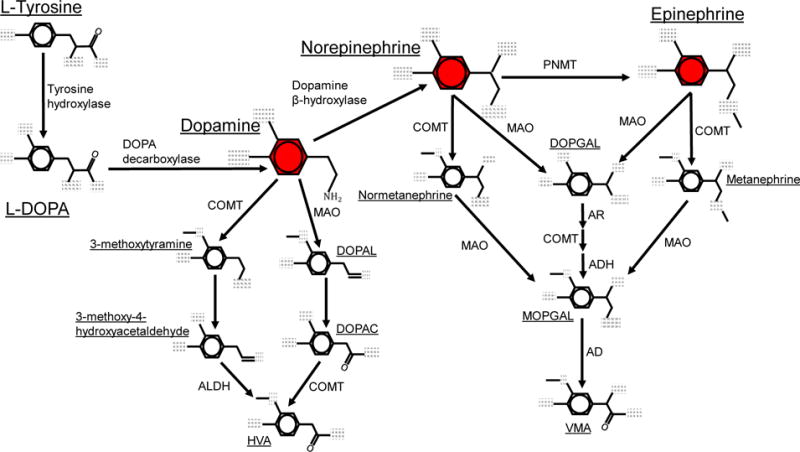
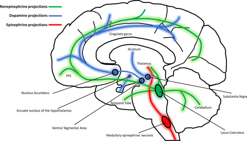
The success of anti-retroviral therapy has improved the quality of life and lifespan of HIV + individuals, transforming HIV infection into a chronic condition. These improvements have come with a cost, as chronic HIV infection and long-term therapy have resulted in the emergence of a number of new pathologies. This includes a variety of the neuropathological and neurocognitive effects collectively known as HIVassociated neurocognitive disorders (HAND) or NeuroHIV. These effects persist even in the absence of viral replication, suggesting that they are mediated the long-term changes in the CNS induced by HIV infection rather than by active replication. Among these effects are significant changes in catecholaminergic neurotransmission, especially in dopaminergic brain regions. In HIV-infected individuals not treated with ARV show prominent neuropathology is common in dopamine-rich brain regions and altered autonomic nervous system activity. Even infected individuals on therapy, there is significant dopaminergic neuropathology, and elevated stress and norepinephrine levels correlate with a decreased effectiveness of antiretroviral drugs. As catecholamines function as immunomodulatory factors, the resultant dysregulation of catecholaminergic tone could substantially alter the development of HIVassociated neuroinflammation and neuropathology. In this review, we discuss the role of catecholamines in the etiology of HIV neuropathogenesis. Providing a comprehensive examination of what is known about these molecules in the context of HIV-associated disease demonstrates the importance of further studies in this area, and may open the door to new therapeutic strategies that specifically ameliorate the effects of catecholaminergic dysregulation on NeuroHIV.

Keywords: Catecholamines; Drug abuse; HIV; HIV-associated neurocognitive disorders; Neuroinflammation.


Figures

Figure 1
Figure 1
Metabolic pathways for catecholamine biosynthesis and degradation. Metabolic pathways for the formation and degradation of dopamine, norepinephrine and epinephrine (shown in red) are described. Catecholamine synthesis is initiated with the hydroxylation of the amino acid tyrosine is by tyrosine hydroxylase (TH), generating L-DOPA. L-DOPA is then converted to dopamine by DOPA decarboxylase (DDC, also known as aromatic L-amino acid decarboxylase, AADC). Dopamine is hydroxylated by dopamine β-hydroxylase (DBH) to form norepinephrine, which is then converted to epinephrine by phenylethanolamine-N-methyltransferase (PNMT). The catecholamines are primarily metabolized by two enzymatic pathways with catechol-O-methyltransferase (COMT) and monoamine oxidase (MAO). COMT converts dopamine to 3-methoxytyramine, norepinephrine to normetanephrine, and epinephrine to metanephrine via meta-O-methylation. MAO converts dopamine to 3,4-Dihydroxyphenylacetaldehyde (DOPAL), and norepinephrine or epinephrine to 3,4-didydroxyphenylclycoaldehyde (DOPGAL) by oxidative deamination. MAO also converts 3-methoxytyramine to 3-methoxy-4-hydroxyacetaldehyde, and the metanephrines to an unstable aldehyde monohydroxyphenylglycol aldehyde (MOPGAL). MOPGAL is ultimately converted to the final product vanillyl mandelic acid (VMA) by aldehyde reductase. In the final steps of dopamine metabolism, COMT and aldehyde dehydrogenase (ALDH) convert DOPAL and 3-methoxy-4-hydroxyacetaldehyde to the final product homovanilic acid (HVA).
Figure 2
Figure 2. Human macrophages express enzymes necessary for the synthesis of norepinephrine and degradation of catecholamines
Human macrophages were generated from peripheral blood mononuclear cells that were isolated from whole blood by ficoll density centrifugation, then matured into monocyte-derived macrophages (MDM) by adherence and maturation for 6 days in 10 ng/mL M-CSF. (A) Expression of mRNA for MAO-A and MAO-B, COMT and DBH is seen in mRNA derived from MDM from 4 donors. (B) Analysis of protein lysates from MDM derived from 3 or 4 additional donors shows expression of MAO (antibody did not distinguish A and B), COMT, and DBH protein.
Figure 3
Figure 3. Anatomical location of catecholaminergic brain regions and projections
Dopamine is predominantly synthesized in midbrain nuclei that give rise to three major dopaminergic projections. The mesocortical pathway arises from the ventral tegmental area (VTA), and ascends via the median forebrain bundle to innervate prefrontal and cingulate cortices to modulate cognition, motivation, and emotional response. The VTA is also the origin of the mesolimbic pathway that projects to the nucleus acumbens, medial PFC, amygdala, hippocampus to mediate reward-based habits, emotions, and cognition. The nigrostriatal pathway ascends from the midbrain substantia nigra pars compacta (SNpc) to striatal nuclei (caudate and putamen) in the forebrain to modulate production and coordination of movement. There is an additional small group of dopamine neurons in the arcuate nucleus of the hypothalamus that project to the median eminence (via the tuberoinfundibular pathway) where dopamine is released into the hypophyseal portal system and transported to the anterior pituitary to inhibit the release of prolactin. Norepinephrine neurons from the locus coeruleus in the brainstem broadly innervate cortical and midbrain regions including the PFC, temporal lobe (hippocampus and amygdala), thalamus, hypothalamus, and cerebellum. Outside the brain, norepinephrine is released as a transmitter from postganglionic sympathetic nerve terminals near the spinal cord, and released as a hormone from the adrenal medulla into the blood stream. The role of norepinephrine is to modulate alertness and arousal (via cortical projections), and autonomic functions (via symphathetic nerves and adrenal medulla) in order to mobilize the body for action. Unlike norepinephrine and dopamine, the majority of epinephrine is synthesized in the periphery, with more than 90% of circulating epinephrine produced by chromaffin cells in the adrenal medulla. Some epinephrine may be synthesized in medullary epinephrine neurons that project to the thalamus and spinal cord, although very little is known about the function of these neurons.
Figure 4
Figure 4
Activation of β-adrenergic receptors does not increase HIV infection in human macrophages. Human macrophages were generated from peripheral blood mononuclear cells that were isolated from whole blood by ficoll density centrifugation, then matured into monocyte-derived macrophages (MDM) by adherence and maturation for 6 days in 10 ng/mL M-CSF. (A) MDM from 5 donors were infected with 10 ng•p24/mL of the brain-derived R5 virus HIVYU2 for 24 hours in the presence (greens) or absence (red) of the isoproterenol (β-adrenergic receptor agonist). Cells were washed after 24 hours and supernatant was collected every 24 hours for 6 days as assayed for p24•Gag as a measure of viral replication. (B) MDM from 5 donors were infected for 2.5 hours with a modified HIVBaL, a lung derived R5 virus, containing an active β-lactamase enzyme, enabling visualization of entry within 9 hours of inoculation. Infections were performed in the absence (red) or presence (greens) of isoproterenol. After 2.5 hours cells were washed to remove excess virus, and incubated in CCF2-AM (ThermoFisher) for 6 hours. After 6 hours, cells were imaged and the percentage of infected cells was enumerated by fluorescent microscopy. Amount of infection displayed as fold change relative to infection with HIV alone, which was set to 1. Statistical analysis for both experiments performed using one-way repeated measures ANOVA.

References

    1. Saylor D, et al. HIV-associated neurocognitive disorder–pathogenesis and prospects for treatment. Nat Rev Neurol. 2016;12(4):234–48. - PMC - PubMed
    1. Heaton RK, et al. HIV-associated neurocognitive disorders before and during the era of combination antiretroviral therapy: differences in rates, nature, and predictors. J Neurovirol. 2011;17(1):3–16. - PMC - PubMed
    1. Rappaport J, Volsky DJ. Role of the macrophage in HIV-associated neurocognitive disorders and other comorbidities in patients on effective antiretroviral treatment. J Neurovirol. 2015;21(3):235–41. - PMC - PubMed
    1. Heaton RK, et al. HIV-associated neurocognitive disorders persist in the era of potent antiretroviral therapy: CHARTER Study. Neurology. 2010;75(23):2087–96. - PMC - PubMed
    1. Woods SP, et al. HIV-associated deficits in action (verb) generation may reflect astrocytosis. J Clin Exp Neuropsychol. 2010;32(5):522–7. - PMC - PubMed

Publication types

MeSH terms