Review
. 2018 Jun;135(6):811-826.
doi: 10.1007/s00401-018-1850-y. Epub 2018 Apr 28.

Somatic mutations in neurons during aging and neurodegeneration

Affiliations
Review

Somatic mutations in neurons during aging and neurodegeneration

Bert M Verheijen et al. Acta Neuropathol. 2018 Jun.

Erratum in

Abstract

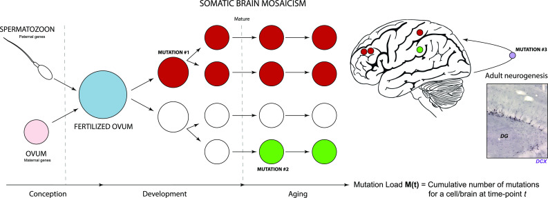
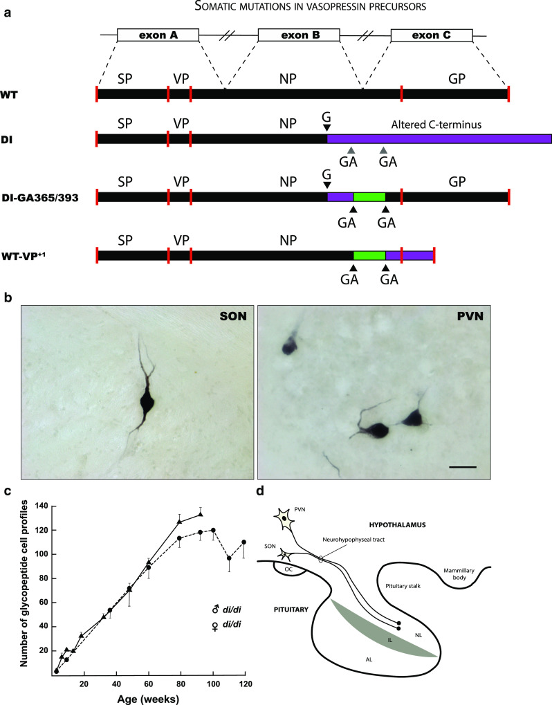
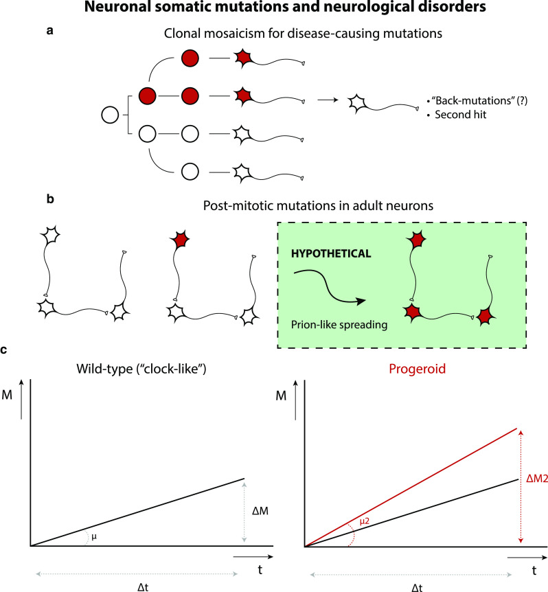
The nervous system is composed of a large variety of neurons with a diverse array of morphological and functional properties. This heterogeneity is essential for the construction and maintenance of a distinct set of neural networks with unique characteristics. Accumulating evidence now indicates that neurons do not only differ at a functional level, but also at the genomic level. These genomic discrepancies seem to be the result of somatic mutations that emerge in nervous tissue during development and aging. Ultimately, these mutations bring about a genetically heterogeneous population of neurons, a phenomenon that is commonly referred to as "somatic brain mosaicism". Improved understanding of the development and consequences of somatic brain mosaicism is crucial to understand the impact of somatic mutations on neuronal function in human aging and disease. Here, we highlight a number of topics related to somatic brain mosaicism, including some early experimental evidence for somatic mutations in post-mitotic neurons of the hypothalamo-neurohypophyseal system. We propose that age-related somatic mutations are particularly interesting, because aging is a major risk factor for a variety of neuronal diseases, including Alzheimer's disease. We highlight potential links between somatic mutations and the development of these diseases and argue that recent advances in single-cell genomics and in vivo physiology have now finally made it possible to dissect the origins and consequences of neuronal mutations in unprecedented detail.

Keywords: Aging; Genome integrity; Neurodegeneration; Neurological disorders; Neuronal development; Somatic brain mosaicism; Somatic mutations.


Conflict of interest statement

The authors declare that the research was conducted in the absence of any commercial or financial relationships that could be construed as a potential conflict of interest.

Figures

Fig. 1
Fig. 1
Somatic mutations in the nervous system. The genome is a set of instructions, or a program, for the development and functioning of organisms. It is often considered to be a fixed chemical entity, which is faithfully copied from mother to daughter cells during successive rounds of cell division and is mostly identical in different cells from different tissues. However, it has become clear that differences in genomes exist between single cells. Some researchers have taken the view that no cell in an individual does, in fact, carry the exact same genetic scripture. This potentially has major implications, especially for post-mitotic cells like neurons that are rarely or not at all replaced during life. Throughout normal development, post-zygotic mutations occur in neural progenitors, which are inherited by their cellular progeny (mutation #1). This will eventually culminate in a genetic mosaic. It has been suggested that somatic mutations can also take place in the developed nervous system (mutation #2; neurogenesis: mutation #3). Mature neurons are generally considered to be terminally differentiated post-mitotic cells with limited regenerative potential. Therefore, they are particularly prone to accumulation of damage. Specifically, genomic integrity of neurons can be influenced by the occurrence of gene mutations. During aging, the nervous system is subjected to various types of stress that contribute to neuronal damage, including genomic alterations such as telomere shortening and chromosomal abnormalities. These changes are accompanied by various other alterations, like impaired nuclear integrity, aberrant nucleocytoplasmic transport and defects at the level of mitochondria (including mtDNA mutations). Insert shows a microscopic image of the dentate gyrus of a mouse hippocampus, immunostained for doublecortin (DCX), a marker for neurogenesis (Verheijen, Vermulst and van Leeuwen, unpublished)
Fig. 2
Fig. 2
Mutations in post-mitotic vasopressin neurons. a Schematic representation of the vasopressin (VP) gene, VP prohormone and mutant forms. The VP gene consists of three exons (exon A, B, and C, consisting of 429 nucleotides), which give rise to a transcript that is spliced to generate the mRNA template for a precursor protein (WT). VP precursor protein is translated in the endoplasmatic reticulum (ER), post-translationally processed and packaged within neurosecretory granules. Subsequently, the protein is axonally transported to nerve terminals in the neural lobe of the pituitary gland (d). In the homozygous (di/di) Brattleboro rat, a single base (G) deletion in exon B results in an out-of-frame protein that contains a poly-lysine tail that cannot be properly processed (DI). This results in (central) diabetes insipidus (DI), a condition that is characterized by polyuria and polydipsia, due to the inability to effectively regulate the VP-mediated retention of water in the kidney’s collection ducts (antidiuretic function). This is an autosomal recessive trait that is inherited in a simple Mendelian fashion. b Intriguingly, some solitary neurons in the supraoptic nucleus (SON) and paraventricular nucleus (PVN) of the homozygous Brattleboro rat appear to be immunoreactive for VP. Bar = 50 μm (Verheijen, Vermulst and van Leeuwen, unpublished). This appears to be due to a second mutation (ΔGA) in a GAGAG motif that is located downstream of the single base mutation (a). As a result the VP mutant precursor (DI-GA365/393) can be processed (i.e., the glycoprotein (GP)-containing part) and the neurosecretory granules can be axonally transported towards the neural lobe. The amount of reverted neurons (+/di) increases age-dependently in both male (filled triangle) and female (filled circle) rats (c). Because GAGAG motifs are also present in the wild-type VP gene of rat and human, a similar process can take place and convert the wild-type VP precursor into an aberrant one (WT-VP+1). This has been shown to occur in hypothalamus of both rat and human. AL anterior lobe, IL intermediate lobe, NL neural lobe, NP neurophysin, OC optic chiasm, SP signal peptide
Fig. 3
Fig. 3
Various types and mechanisms of neuronal somatic mutations. Different types of mutations have been found to be present in single neurons, including long interspersed nuclear element 1 (L1 or LINE1) retrotransposition, copy-number variations (CNVs), single-nucleotide variants (SNVs), and microsatellite/short tandem repeat variants. The exact contribution of each of these events to somatic neuronal mosaicism is unknown. Also, the mechanisms through which these mutations can arise are mostly unknown. Slippage of DNA polymerases, e.g., due to secondary structures in the chromatin, can cause changes in length of microsatellites. Cytosine deamination has been recognized as a frequent cause of SNVs. It will be necessary to accurately quantify these different types of mutations, for various cell types and brain regions, during different developmental stages or under particular conditions, in order to gain insight into their (potential) roles
Fig. 4
Fig. 4
Neuronal somatic mutations and neurological disorders. Somatic mutations in neurons could cause or predispose for neuronal diseases. Neuronal somatic mutations can either occur in neuronal progenitors, giving rise to mutant daughter cells through clonal expansion of these mutation-carrying cells (a) or in post-mitotic neurons (b), resulting in very fine changes in the nervous system. A combination of both might reflect an intriguing mechanistic link between developmental and degenerative brain disorders. Neuron-to-neuron spreading of pathological proteins could provide an explanation for widespread pathology induced by single-neuron DNA mutations, but this model is purely hypothetical as of now. c Somatic mutations have been found to accumulate in neurons as a function of age. Neuronal somatic mutation accumulation could be a general hallmark of neuronal aging. In DNA repair disorders (Cockayne syndrome, Xeroderma pigmentosum) increased rates of somatic mutations (μ) have been observed (“progeroid” phenotype). Increased rates of somatic mutation in single neurons could be important for other neurodegenerative disorders and may also accelerate neuronal aging itself
Fig. 5
Fig. 5
Experimental strategies to model brain somatic mosaicism. Different experimental model systems can be used to mimic somatic mosaicism in the nervous system. For in vitro study of neuronal somatic mutations, hiPSC-derived neurons would be an attractive option, because they are human cells and can be derived from little starting material (through clonal expansion). Brain organoids are 3-dimensional models for neuronal brain development and disease. These also allow grafting of single neurons and grafting into live brains or other parts of organoids (e.g., to study mechanisms of prion-like spreading). Manipulated organoids can be optically cleared and imaged in 3D, e.g., using light sheet microscopy. Depending on the biological questions being posed, and especially the window of time and spatial requirements, different approaches can be considered. To study the in vivo contribution of somatic mutations, gene-editing constructs can be transfected in developing mouse brain via in utero electroporation of embryonic mice. Electroporation of a mouse brain (or organoid) will result in mosaic expression by default, because only select cells are transfected. In the adult brain, delivery of gene-editing ribonucleoprotein complexes (RNPs) or viral agents can be considered. Alternatively, (inducible) transgenic mice can be used to generate mosaics (Verheijen, Vermulst and van Leeuwen, unpublished)
Fig. 6
Fig. 6
Neuronal epimutations. a Schematic representation of “molecular misreading”, a form of transcriptional mutagenesis or “epimutation”. (Epi)genomic drift and an overall increase in transcriptional noise may contribute to age-related neurodegenerative processes. Dinucleotide deletions (e.g., ΔGA or ΔGU) in an mRNA molecule can result in the generation of mutant proteins, which can be recognized by specific antibodies. b An example of such a mutant protein is ubiquitin-B+1 (UBB+1). UBB+1 is detectable in the brains of Alzheimer’s disease (AD) patient brains, by immunocytochemistry using antibodies that specifically recognize the abnormal C-terminal domain of UBB+1. This demonstrates the importance of neuropathological observation in validating these mutational events. Scale bar: 200 μm. c Transgenic expression of UBB+1 in mice results in behavioral phenotypes that are consistent with neurodegeneration. Image shows a sagittal section of the hippocampus of a UBB+1 transgenic mouse, stained for UBB+1. Scale bar: 200 μm (Verheijen, Vermulst and van Leeuwen, unpublished)

References

    1. Aguzzi A, Lakkaraju AKK. Cell biology of prions and prionoids: a status report. Trends Cell Biol. 2016;26:40–51. doi: 10.1016/j.tcb.2015.08.007. - DOI - PubMed
    1. Alexandrov LB, Jones PH, Wedge DC, Sale JE, Campbell PJ, Nik-Zainal S, et al. Clock-like mutational processes in human somatic cells. Nat Genet. 2015;47:1402–1407. doi: 10.1038/ng.3441. - DOI - PMC - PubMed
    1. Alzualde A, Moreno F, Martínez-Lage P, Ferrer I, Gorostidi A, Otaegui D, et al. Somatic mosaicism in a case of apparently sporadic Creutzfeldt-Jakob disease carrying a de novo D178 N mutation in the PRNP gene. Am J Med Genet B Neuropsychiatr Genet. 2010;153B:1283–1291. doi: 10.1002/ajmg.b.31099. - DOI - PubMed
    1. Amunts K, Zilles K. Architectonic mapping of the human brain beyond brodmann. Neuron. 2015;88:1086–1107. doi: 10.1016/j.neuron.2015.12.001. - DOI - PubMed
    1. Ando K, Laborde Q, Lazar A, Godefroy D, Youssef I, Amar M, et al. Inside Alzheimer brain with CLARITY: senile plaques, neurofibrillary tangles and axons in 3-D. Acta Neuropathol. 2014;128:457–459. doi: 10.1007/s00401-014-1322-y. - DOI - PMC - PubMed

Publication types