. 2018 May 3;102(5):832-844.
doi: 10.1016/j.ajhg.2018.03.013. Epub 2018 Apr 26.

Monoallelic Mutations to DNAJB11 Cause Atypical Autosomal-Dominant Polycystic Kidney Disease

Affiliations

Monoallelic Mutations to DNAJB11 Cause Atypical Autosomal-Dominant Polycystic Kidney Disease

Emilie Cornec-Le Gall et al. Am J Hum Genet. .

Abstract

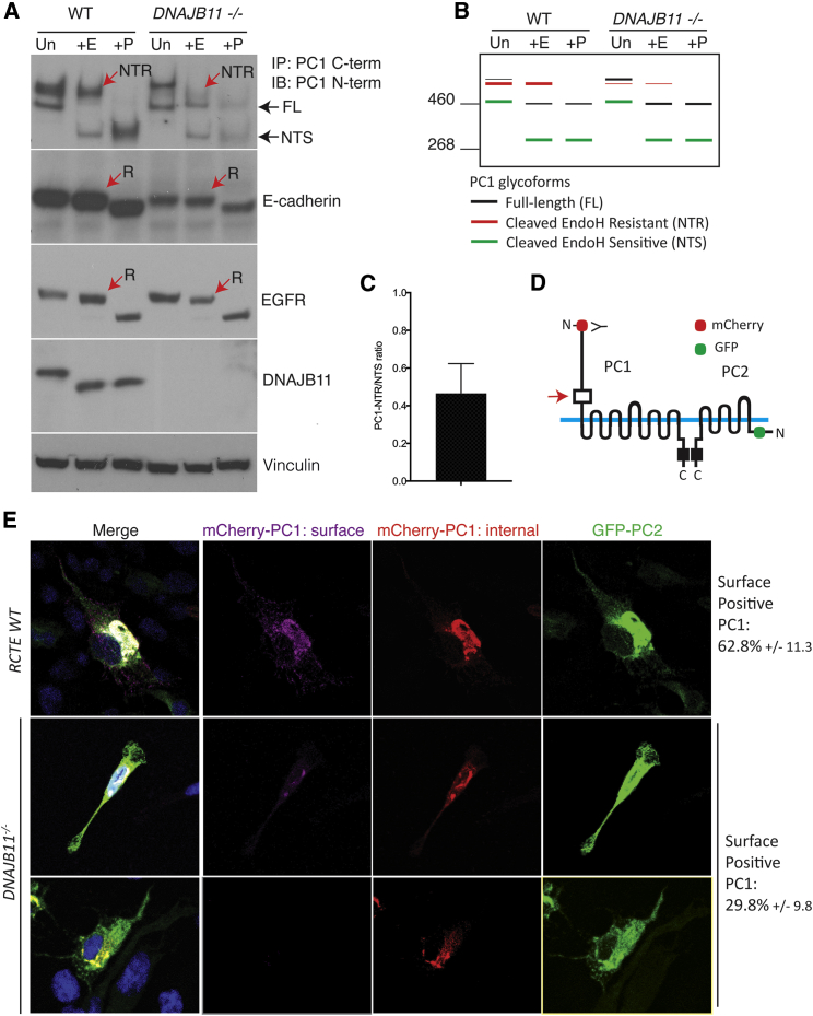
Autosomal-dominant polycystic kidney disease (ADPKD) is characterized by the progressive development of kidney cysts, often resulting in end-stage renal disease (ESRD). This disorder is genetically heterogeneous with ∼7% of families genetically unresolved. We performed whole-exome sequencing (WES) in two multiplex ADPKD-like pedigrees, and we analyzed a further 591 genetically unresolved, phenotypically similar families by targeted next-generation sequencing of 65 candidate genes. WES identified a DNAJB11 missense variant (p.Pro54Arg) in two family members presenting with non-enlarged polycystic kidneys and a frameshifting change (c.166_167insTT) in a second family with small renal and liver cysts. DNAJB11 is a co-factor of BiP, a key chaperone in the endoplasmic reticulum controlling folding, trafficking, and degradation of secreted and membrane proteins. Five additional multigenerational families carrying DNAJB11 mutations were identified by the targeted analysis. The clinical phenotype was consistent in the 23 affected members, with non-enlarged cystic kidneys that often evolved to kidney atrophy; 7 subjects reached ESRD from 59 to 89 years. The lack of kidney enlargement, histologically evident interstitial fibrosis in non-cystic parenchyma, and recurring episodes of gout (one family) suggested partial phenotypic overlap with autosomal-dominant tubulointerstitial diseases (ADTKD). Characterization of DNAJB11-null cells and kidney samples from affected individuals revealed a pathogenesis associated with maturation and trafficking defects involving the ADPKD protein, PC1, and ADTKD proteins, such as UMOD. DNAJB11-associated disease is a phenotypic hybrid of ADPKD and ADTKD, characterized by normal-sized cystic kidneys and progressive interstitial fibrosis resulting in late-onset ESRD.

Keywords: ADPKD; ADPLD; ADTKD; DNAJB11; pathogenic variants; renal cystic disease.


Figures

Figure 1
Figure 1
Pedigrees of the Seven Families with DNAJB11-Associated Disorder, Abdominal Imaging, and Histological Characteristics (A) Seven pedigrees: black squares or circles indicate affected male or female subjects, respectively, presenting with bilateral renal cysts, liver cysts, renal failure, and/or genetically diagnosed, and gray symbols indicate case subjects where clinical information is unavailable. Clinical characteristics are detailed in Table 1. The DNAJB11 pathogenic variants identified in each family are indicated below each pedigree, “Mut” denotes that DNA sequencing proved the mutation, “WT” (wild-type) denotes the absence of the pathogenic variant, and “ND” (no data) indicates that no DNA sample was available. The DNAJB11-affected families originate from the US (families 1 [M624], 3 [M775], and 5 [M1231]), Canada (family 2 [YU209]), Belgium (family 4 [M1122]), and France (families 6 [PK20211] and 7 [PK4114]). Families came from various study cohorts: HALT-PKD (M775), CRISP (M1231), GeneQuest (NCT02112136, PK20211), TGESP (YU209), the French National Institute of Health and Medical Research Unit 1078, Brest, France (PK4114), and the Mayo PKD Center (M624, M1122). (B–J) Abdominal imaging of nine individuals from the seven families. Seven have magnetic resonance imaging (MRI; T2-weighted in B–E, G, J; T1-weighted in H) and two computed tomography (CT; non contrast enhanced in F; contrast enhanced in I). (K) Two representative kidney histology sections of family 5, II.2, who underwent a bilateral nephrectomy, stained with Masson’s trichrome (blue indicates fibrosis).
Figure 2
Figure 2
Identification of DNAJB11 Pathogenic Variants in the Seven Pedigrees (A) Integrated Genomics Viewer (IGV, Broad Institute) visualization of the two DNAJB11 changes independently identified by WES (sequences from family 1, II.1 and family 2, III.2), with details of read depths and allele frequencies tabulated for each WES-identified pathogenic change. Targeted next-generation sequencing led to the identification of three additional pathogenic variants in five different pedigrees. (B) Sanger confirmations of the five identified DNAJB11 changes; WT sequences are shown for comparison. (C) Domain organization of DNAJB11, with the distribution of the pathogenic variants identified. DNAJB11 is a 358-amino-acid protein comprising a highly conserved J domain, with a characteristic His-Pro-Asp (HPD) motif through which it interacts with the chaperone BiP, a substrate binding domain, and a dimerization domain. The two identified missense changes occurred in the J-domain, one of them (p.Pro54Arg, identified in family 1) affecting the HPD motif. The other variants, all truncating, occurred in the J-domain or the substrate binding domain of the protein. (D) Multiple sequence alignments of DNAJB11 orthologous proteins, from human to rice (top), and of the J-domains of other human proteins from the ERdj family (bottom). The two amino acids affected by missense changes, proline at position 54 and leucine at position 77, are invariant in both cases.
Figure 3
Figure 3
Loss of DNAJB11 Affects Maturation and Localization of Polycystin-1 (A) N-linked deglycosylation analysis of WT and DNAJB11−/− RCTE membrane proteins either untreated (Un) or treated with Endoglycosidase H (+E; removing immature glycans) or PNGaseF (+P; removing all sugars). Immunoprecipitation was used to enrich the PC1 complex with a C-terminal PC1 (PC1-CT) antibody and immunodetected with the N-terminal PC1 (PC1-NT) antibody. Partial loss of mature PC1 (N-terminal PC1, resistant to EndoH, abbreviated NTR and indicated by a red arrow) was observed in DNAJB11-deficient cells (see C for quantification), and increased levels of immature PC1 (including full-length [FL] and N-terminal EndoH sensitive [NTS] PC1). Similar analysis of E-cadherin and epidermal growth factor receptor (EGFR) did not show altered sensitivity to maturation in DNAJB11−/− cells, although higher levels of E-cadherin were observed in wild-type as compared to DNAJB11−/− cells. DNAJB11 loss was confirmed in null cells, and vinculin was employed as a loading control. (B) Schematic representation of the PC1 products in WT and DNAJB11−/− cells presented in (A). (C) Decreased ratio of PC1 NTR compared to PC1 NTS in DNAJB11−/− cells by more than 50%. The PC1 NTR/NTS bands density were measured in three different DNAJB11−/− clones and three replicates of one clone, normalized to WT results, and presented here as the mean and standard deviation of all experiments. (D) Diagram of the WT-tagged PC1 and PC2 constructs used for the localization analysis presented in (E) with the site of PC1 GPS cleavage indicted with an arrow. (E) WT or DNAJB11−/− cells were transfected with mCherry-PC1 and GFP-tagged-PC2 and examined for surface PC1 labeling (mCherry) in co-transfected cells. Surface PC1 expression was observed in 62.8% of the co-transfected WT cells, but only in 29.8% of the co-transfected DNAJB11−/− (p < 0.001). This ∼50% decrease of surface PC1 expression in DNAJB11−/− as compared to WT cells was confirmed on three different experiments. DNAJB11−/− cells positive (middle) and negative (bottom) for surface PC1 are shown.
Figure 4
Figure 4
Intracellular Distribution of UMOD, BiP, and MUC1 in Tubular Epithelial Cells Immunofluorescence labeling of uromodulin (UMOD; top), MUC1 (middle), and BiP (bottom) in kidney from an unaffected individual (control) and two DNAJB11-affected individuals (family 1, II.1 and family 5, II.2). The UMOD expression in the tubular ascending loop of Henle (TAL) of the unaffected individual is apical but appears to be retained intracellularly in the two DNAJB11-affected individuals (see insets a–c). Cellular distribution of BiP and MUC1 are not markedly different in the control and affected individuals, although in family 5, II.2 several TAL cells have apparent intracellular retention of MUC1 (yellow arrows) with increased intensity of BiP staining (red arrows; insets d and e).

Comment in

References

    1. Ong A.C., Devuyst O., Knebelmann B., Walz G., Diseases, ERA-EDTA Working Group for Inherited Kidney Diseases Autosomal dominant polycystic kidney disease: the changing face of clinical management. Lancet. 2015;385:1993–2002. - PubMed
    1. The European Polycystic Kidney Disease Consortium The polycystic kidney disease 1 gene encodes a 14 kb transcript and lies within a duplicated region on chromosome 16. Cell. 1994;77:881–894. - PubMed
    1. Mochizuki T., Wu G., Hayashi T., Xenophontos S.L., Veldhuisen B., Saris J.J., Reynolds D.M., Cai Y., Gabow P.A., Pierides A. PKD2, a gene for polycystic kidney disease that encodes an integral membrane protein. Science. 1996;272:1339–1342. - PubMed
    1. Heyer C.M., Sundsbak J.L., Abebe K.Z., Chapman A.B., Torres V.E., Grantham J.J., Bae K.T., Schrier R.W., Perrone R.D., Braun W.E., HALT PKD and CRISP Investigators Predicted mutation strength of nontruncating PKD1 mutations aids genotype-phenotype correlations in autosomal dominant polycystic kidney disease. J. Am. Soc. Nephrol. 2016;27:2872–2884. - PMC - PubMed
    1. Cornec-Le Gall E., Audrézet M.P., Chen J.M., Hourmant M., Morin M.P., Perrichot R., Charasse C., Whebe B., Renaudineau E., Jousset P. Type of PKD1 mutation influences renal outcome in ADPKD. J. Am. Soc. Nephrol. 2013;24:1006–1013. - PMC - PubMed

Publication types

MeSH terms