

doi: 10.1055/s-0037-1607047.
Epub 2017 Nov 27.
"The Serpentine Zone": A Surgeon's Guide to the Surface Anatomy of the Digital Neurovascular Spiral in Dupuytren's Contracture
Affiliations
- PMID: 29706740
- PMCID: PMC5919782
- DOI: 10.1055/s-0037-1607047
Item in Clipboard
"The Serpentine Zone": A Surgeon's Guide to the Surface Anatomy of the Digital Neurovascular Spiral in Dupuytren's Contracture
J Hand Microsurg.
2018 Apr.
Abstract
The anatomy of the cords that form in Dupuytren's disease is complicated and a spiral cord is the most challenging variant to address. It displaces the neurovascular bundle toward or beyond the midline and closer to the skin. This article illustrates the surface anatomy of the neurovascular spiral to help surgeons identify this zone of danger that the authors term "the serpentine zone." Careful dissection in this zone will help avoid iatrogenic digital neurovascular injury.
Keywords: Dupuytren's; spiral cords; surface anatomy.
Conflict of interest statement
Figures
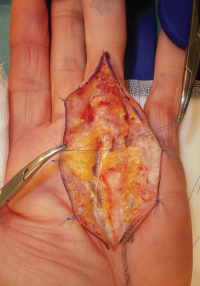
An intraoperative picture showing the radial spiral cord with a serpentine neurovascular bundle, deviated to the ulnar side, around which a suture loop has been passed for identification. The ulnar neurovascular bundle is also shown with a suture loop.

Surface anatomy of the serpentine radial digital neurovascular bundle represented by the solid line from the radial side toward the midline incision. The arrow indicates the level at which the nerve was most deviated and superficial in this patient. The proximal extent of the serpentine zone is the distal palmar crease and the distal extent is the proximal finger crease.

Both neurovascular bundles have returned to their normal position and are parallel to each other following fasciectomy.

Wound closure using Z-plasties.

(
A
,
B
) Postoperative results showing full extension and flexion of the digit and good wound healing.
References
-
- Brunton L M, Chhabra B. 7th ed. Philadelphia, PA: Elsevier; 2016. Hand, upper extremity and microvascular surgery; pp. 626–630.
-
- McFarlane R M. Patterns of the diseased fascia in the fingers in Dupuytren's contracture. Displacement of the neurovascular bundle. Plast Reconstr Surg. 1974;54(01):31–44. - PubMed
-
- McGrouther D A. Edinburgh, Scotland: Churchill Livingstone; 1990. Dupuytren's Disease; pp. 293–312.
LinkOut - more resources
Full Text Sources
Other Literature Sources
