. 2018 Apr 13:12:13.
doi: 10.3389/fnsys.2018.00013. eCollection 2018.

Inflammation and Tissue Remodeling in the Bladder and Urethra in Feline Interstitial Cystitis

Affiliations

Inflammation and Tissue Remodeling in the Bladder and Urethra in Feline Interstitial Cystitis

F Aura Kullmann et al. Front Syst Neurosci. .

Erratum in

Abstract

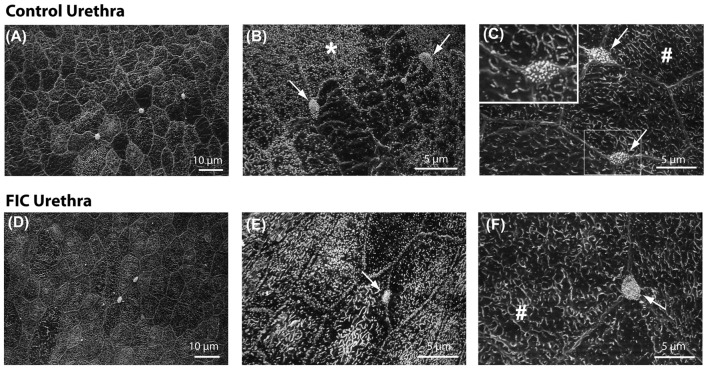
Interstitial cystitis/bladder pain syndrome (IC/BPS) is a debilitating chronic disease of unknown etiology. A naturally occurring disease termed feline interstitial cystitis (FIC) reproduces many features of IC/BPS patients. To gain insights into mechanisms underlying IC/BPS, we investigated pathological changes in the lamina propria (LP) of the bladder and proximal urethra in cats with FIC, using histological and molecular methods. Compared to control cat tissue, we found an increased number of de-granulated mast cells, accumulation of leukocytes, increased cyclooxygenase (COX)-1 expression in the bladder LP, and increased COX-2 expression in the urethra LP from cats with FIC. We also found increased suburothelial proliferation, evidenced by mucosal von Brunn's nests, neovascularization and alterations in elastin content. Scanning electron microscopy revealed normal appearance of the superficial urethral epithelium, including the neuroendocrine cells (termed paraneurons), in FIC urethrae. Together, these histological findings suggest the presence of chronic inflammation of unknown origin leading to tissue remodeling. Since the mucosa functions as part of a "sensory network" and urothelial cells, nerves and other cells in the LP are influenced by the composition of the underlying tissues including the vasculature, the changes observed in the present study may alter the communication of sensory information between different cellular components. This type of mucosal signaling can also extend to the urethra, where recent evidence has revealed that the urethral epithelium is likely to be part of a signaling system involving paraneurons and sensory nerves. Taken together, our data suggest a more prominent role for chronic inflammation and tissue remodeling than previously thought, which may result in alterations in mucosal signaling within the urinary bladder and proximal urethra that may contribute to altered sensations and pain in cats and humans with this syndrome.

Keywords: bladder; paraneurons; urethra; urothelium; von Brunn’s nest.


Figures

Figure 1
Figure 1
Age and sex distribution of control (C) and feline interstitial cystitis (FIC) cats. Open circle symbols indicate males, closed circle symbols indicate females. Statistical significance was tested using unpaired t-test. Asterisk (*) indicates p < 0.05.
Figure 2
Figure 2
Mast cell quantification in bladder tissue from control and FIC cats. (A) Example of de-granulated mast cells, (red arrows) from a FIC cat. (Ai) Inset shows higher magnification of the area indicated by the red arrows and illustrates the presence of de-granulated mast cells in the vicinity of a blood vessel (red star placed in the lumen of the blood vessel). (B,C) Examples of de-granulated (B) and not de-granulated (C) mast cells from an FIC cat. (D–I) Quantification of mast cells and de-granulated (dg-) mast cells in bladder tissue from control (n = 17; individual data points shown in black, and average ± SEM in gray) and FIC (n = 13; individual data points shown in gray, and average ± SEM in black) cats. Statistical significance was tested using Mann-Whitney test. Asterisk (*) indicates p < 0.05. (D) Total number of mast cells, including granulated and de-granulated, in bladder tissue which is comprised of lamina propria (LP) and smooth muscle (SM) in control and FIC tissue (p = 0.50). (E) Total number of de-granulated mast cells in the bladder in control and FIC tissue (p = 0.037). (F) Total number of mast cells in the LP in control and FIC tissue (p = 0.38). (G) Total number of de-granulated mast cells in the LP in control and FIC tissue (p = 0.01). (H) Total number of mast cells in the SM in control and FIC tissue (p = 0.90). (I) Total number of de-granulated mast cells in the SM in control and FIC tissue (p = 0.47). In all panels, each symbol represents data from one animal. Open circles represent males, closed circles females and open squares represent undocumented sex. In (C), the highest data point is from a male FIC cat 48 months old that fit the criteria to be included in the study.
Figure 3
Figure 3
Mast cell quantification in urethral tissue from control and FIC cats. Data are presented as single points from individual cats with mean ± SEM. Control cats (n = 3) are shown in black and FIC (n = 7) in gray. Statistical significance was tested using Mann-Whitney test. (A) Total number of mast cells, including granulated and de-granulated (dg-), in the urethral tissue which is comprised of LP and SM in control and FIC tissue (p = 0.18). (B) Total number of de-granulated mast cells in the whole urethra in control and FIC tissue (p = 0.60). (C) Total number of mast cells in the LP in control and FIC tissue (p = 0.11). (D) Total number of de-granulated mast cells in the LP in control and FIC tissue (p = 0.55). (E) Total number of mast cells in the SM in control and FIC tissue (p = 0.18). (F) Total number of de-granulated mast cells in the SM in control and FIC tissue (p = 0.16). In all panels, each symbol represents data from one animal. Open circles represent males, closed circles females and open squares represent undocumented sex.
Figure 4
Figure 4
Immune cell infiltration in the LP of the bladder and urethra. (A) Example of leukocyte accumulation and area (red line) used for quantification, in the bladder of a FIC cat. Inset illustrates a neutrophil, characterized by the presence of dark small vesicles. (B,C) Histogram of area of immune cell infiltrate in the LP of the bladder from control (black; n = 17) and FIC (gray; n = 13) cats (Kolmogorov-Smirnov test, p = 0.02) cats showing only values smaller than 6000 μm2. One measurement in control and two measurements in FIC tissue were larger than 6000 μm2. Bin size 200 μm2. (D,E) Histogram of area of immune cell infiltrate in the LP of the urethra of control (black; n = 3) and FIC (gray; n = 7) cats (Kolmogorov-Smirnov test, p = 0.02) showing only values smaller than 3500 μm2. Bin size 200 μm2. There was no immune cell infiltrate in the urethra of control cats (black bar centered at 0; n = 3 cats). Three measurements in FIC tissue were larger than 3500 μm2. Statistical significance was tested using Kolmogorov-Smirnov test. Asterisk (*) indicates p < 0.05.
Figure 5
Figure 5
Cyclooxygenase (COX)-1 and COX-2 expression in the mucosa of the bladder and urethra. (A) COX-1 expression in the mucosa of the bladder (circles) and urethra (triangles) from control (n = 13 bladders and 12 urethral tissues) and FIC (n = 20 bladder and 14 urethral tissues) cats. Statistical significance was tested using Mann-Whitney test (bladder p = 0.03, urethra p = 0.31). (B) COX-2 expression in the mucosa of the bladder (circles) and urethra (triangles) from control (n = 13 bladders and 12 urethral tissues) and FIC (n = 20 bladder and 12 urethral tissues) cats. Statistical significance was tested using Mann-Whitney test (bladder p = 0.47, urethra p = 0.047). Data are presented as single points from individual cats with mean ± SEM. Asterisk (*) indicates p < 0.05. In all panels, each symbol represents data from one animal. Open circles represent males, closed circles females and open squares represent undocumented sex. (C) Representative blots from bladder and urethra indicating the expression of COX-1 and COX-2. Each of the three lines (control and FIC) represents tissue from a single cat probed for COX-1 (upper panel) COX-2 middle panel, and total protein (lower panel) which was used for normalization. The expected band sizes for COX-1 and COX-2 are 75 kDa.
Figure 6
Figure 6
Von Brunn’s nests in the bladder and urethra. (A) Example of von Brunn’s nests indicated by yellow arrows, in the bladder of an FIC cat. (B) Example of von Brunn’s nests (inside the yellow circle) in the urethra of a FIC cat. (C) Percentage of control (black) and FIC (gray) cats presenting von Brunn’s nests in the bladder and urethra. Numbers on bar graphs represent the number of cats exhibiting von Brunn’s nests and the total number of cats assessed. Statistical significance was tested using Chi-square test: p = 0.02 for bladder and p = 0.17 for urethra. Asterisk (*) indicates p < 0.05.
Figure 7
Figure 7
Increased angiogenesis in LP of the bladder and urethra. (A,B) Example of blood vessels in Mason trichrome stained tissues from control (A) and FIC (B) cat bladder. Insets (Ai,Bi) are magnified images of the areas outlined in yellow in (A,B), to illustrate blood vessels (indicated by yellow stars placed next to the vessels). (C,D) Number of blood vessels per area of tissue in the bladder (C) and urethra (D) from control (black; n = 17 bladder and n = 3 urethral tissues; Mann-Whitney test p = 0.024) and FIC (gray; n = 13 bladder and n = 7 urethral tissues; Mann-Whitney test p = 0.3) cats. Each symbol represents data from one animal. Open circles represent males, closed circles females and open squares represent undocumented sex. Statistical significance was tested using Mann-Whitney test. Asterisk (*) indicates p < 0.05.
Figure 8
Figure 8
Increased elastin expression in the LP of the bladder and urethra. (A,B) Example of elastin fibers detected by Verhoeff-Van Gieson staining, in the bladder from control (A) and FIC (B) cats. (C,D) High magnification of the areas outlined in yellow in (A,B). Note black fibers are more prominent in the FIC tissue (B,D). (E,F) Elastin fiber density (quantified as number of fibers per tissue area) in the bladder (E) and urethra (F). Data are from 16 bladders and three urethrae from control cats shown in black, and 12 bladders and seven urethrae from FIC cats shown in gray. Each symbol represents data from one animal. Open circles represent males, closed circles females and open squares represent undocumented sex. Statistical significance was evaluated using Mann-Whitney test. Asterisks (*) indicate p < 0.05 (p = 0.03 and p = 0.01 for bladder and urethra, respectively).
Figure 9
Figure 9
Urethral epithelial structure. (A–F) Scanning electron microscopy representative images of proximal urethral epithelium from control (A–C) and FIC (D–F) cats. (A,D) Low magnification views of representative areas. (B,E) High magnification areas from control (B) and FIC (E), illustrating cells covered by short microvilli, indicated by *. (C,F) High magnification areas from control (C) and FIC (F) illustrating cells similar to bladder umbrella cells, indicated by #. Arrows in (B,C,E,F), and inset in (C) illustrate paraneurons, characterized by a tuft of microvilli-like appearance and located at the intersection of large polygonal cells (inset C).

References

    1. Ackerman A. L., Jellison F. C., Lee U. J., Bradesi S., Rodríguez L. V. (2016). The Glt1 glutamate receptor mediates the establishment and perpetuation of chronic visceral pain in an animal model of stress-induced bladder hyperalgesia. Am. J. Physiol. Renal Physiol. 310, F628–F636. 10.1152/ajprenal.00297.2015 - DOI - PMC - PubMed
    1. Aizawa N., Igawa Y., Nishizawa O., Wyndaele J. J. (2010). Effects of CL316,243, a β 3-adrenoceptor agonist and intravesical prostaglandin E2 on the primary bladder afferent activity of the rat. Neurourol. Urodyn. 29, 771–776. 10.1002/nau.20826 - DOI - PubMed
    1. Barry C. M., Ji E., Sharma H., Yap P., Spencer N. J., Matusica D., et al. . (2017). Peptidergic nerve fibers in the urethra: morphological and neurochemical characteristics in female mice of reproductive age. Neurourol. Urodyn. [Epub ahead of print]. 10.1002/nau.23434 - DOI - PubMed
    1. Birder L. A., Barrick S. R., Roppolo J. R., Kanai A. J., de Groat W. C., Kiss S., et al. . (2003). Feline interstitial cystitis results in mechanical hypersensitivity and altered ATP release from bladder urothelium. Am. J. Physiol. Renal Physiol. 285, F423–F429. 10.1152/ajprenal.00056.2003 - DOI - PubMed
    1. Birder L. A., de Wachter S., Gillespie J., Wyndaele J. J. (2014). Urethral sensation: basic mechanisms and clinical expressions. Int. J. Urol. 21, 13–16. 10.1111/iju.12349 - DOI - PubMed