. 2018 Apr 12:9:732.
doi: 10.3389/fmicb.2018.00732. eCollection 2018.

The higBA Toxin-Antitoxin Module From the Opportunistic Pathogen Acinetobacter baumannii - Regulation, Activity, and Evolution

Affiliations

The higBA Toxin-Antitoxin Module From the Opportunistic Pathogen Acinetobacter baumannii - Regulation, Activity, and Evolution

Julija Armalytė et al. Front Microbiol. .

Abstract

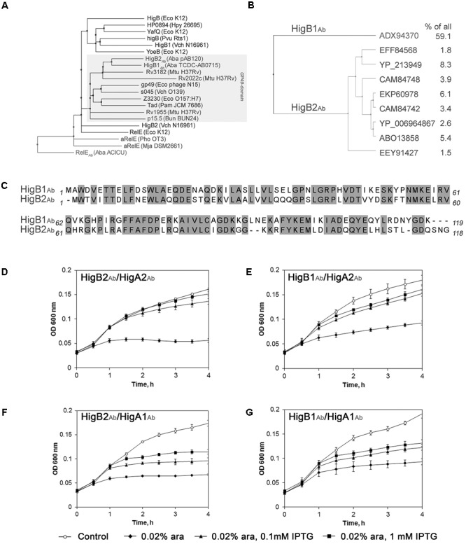
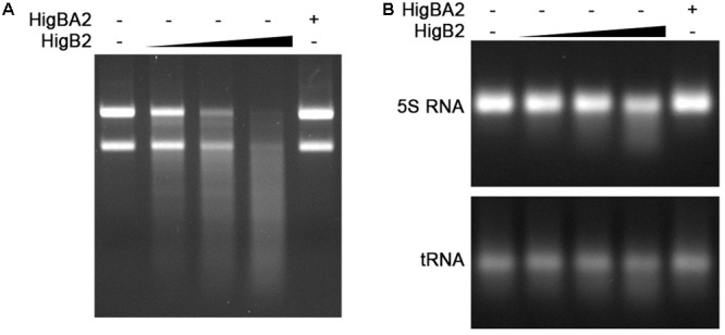
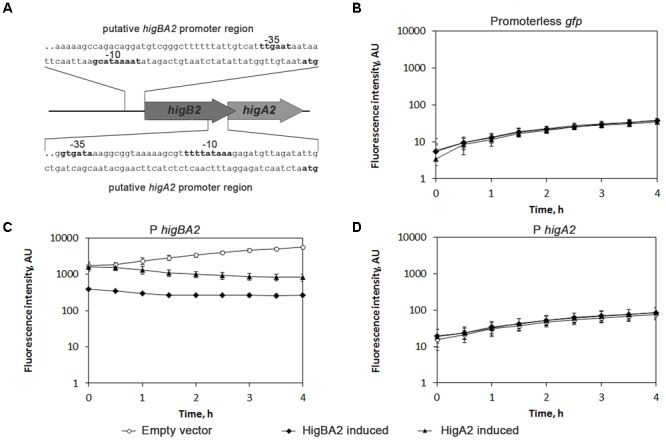
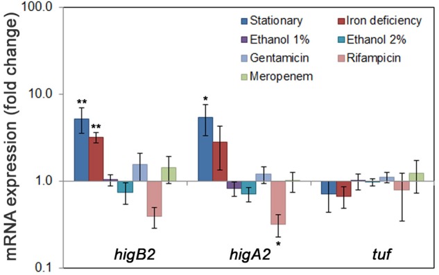
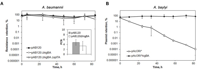
Acinetobacter baumannii is one of the major causes of hard to treat multidrug-resistant hospital infections. A. baumannii features contributing to its spread and persistence in clinical environment are only beginning to be explored. Bacterial toxin-antitoxin (TA) systems are genetic loci shown to be involved in plasmid maintenance and proposed to function as components of stress response networks. Here we present a thorough characterization of type II system of A. baumannii, which is the most ubiquitous TA module present in A. baumannii plasmids. higBA of A. baumannii is a reverse TA (the toxin gene is the first in the operon) and shows little homology to other TA systems of RelE superfamily. It is represented by two variants, which both are functional albeit exhibit strong difference in sequence conservation. The higBA2 operon is found on ubiquitous 11 Kb pAB120 plasmid, conferring carbapenem resistance to clinical A. baumannii isolates and represents a higBA variant that can be found with multiple sequence variations. We show here that higBA2 is capable to confer maintenance of unstable plasmid in Acinetobacter species. HigB2 toxin functions as a ribonuclease and its activity is neutralized by HigA2 antitoxin through formation of an unusually large heterooligomeric complex. Based on the in vivo expression analysis of gfp reporter gene we propose that HigA2 antitoxin and HigBA2 protein complex bind the higBA2 promoter region to downregulate its transcription. We also demonstrate that higBA2 is a stress responsive locus, whose transcription changes in conditions encountered by A. baumannii in clinical environment and within the host. We show elevated expression of higBA2 during stationary phase, under iron deficiency and downregulated expression after antibiotic (rifampicin) treatment.

Keywords: Acinetobacter baumannii; HigBA; plasmid maintenance; protein complex; toxin-antitoxin.


Figures

FIGURE 1
FIGURE 1
Phylogenetic and homology analysis of Acinetobacter baumannii HigBAb. (A) Cluster of GP49-domain toxin family within RelE superfamily, represented as neighbor joining tree. Gray box covers all GP49-domain proteins. The full organism names are provided in the Supplementary Material (Supplementary Table S3); (B) Two major lineages of HigBAb toxins, represented as average distance tree. All proteins that have more than 1% identical hits in all deposited sequences are shown and their frequency in percentage is provided; (C) Alignment of pAB120-borne HigB2Ab (Povilonis et al., 2013) and HigB1Ab (Jurenaite et al., 2013) toxin sequences. Identical amino acid residues are outlined in gray boxes. All alignments were prepared as described in Section “Materials and Methods”; (D–G) Growth of Escherichia coli BW25113F′ harboring components of higBAAb and their combinations. The E. coli strains with pUHE-“antitoxin” and pBAD30-“toxin” plasmids were grown until OD600 = 0.1 and induced with 0.02% arabinose and/or with 0.1–1 mM IPTG as described in Section “Materials and Methods.” The data from three independent experiments are represented, the error bars indicate standard deviation.
FIGURE 2
FIGURE 2
Analysis of HigB2Ab–HigA2Ab protein interaction. (A) Two-hybrid analysis of interaction of toxin-antitoxin (TA) components. E. coli BTH101 with two plasmids, one encoding Cya-T18 fused to A. baumanii antitoxins HigA2Ab or RelBAb, and another encoding Cya-T25 fused to toxins HigB2Ab or RelEAb. The ability of the proteins to interact and reconstitute functional Cya from T25 and T18 was observed as blue colony formation when grown on LB agar with IPTG and X-gal for 24 h; (B) Size-exclusion chromatography and SDS–PAGE analysis of His-HigBA2 and HigBA2-His protein complexes. The proteins used for molecular mass standard curve are indicated as black diamonds, empty diamonds indicate the positions of His-HigBA2 and HigBA2-His protein complexes and single His-HigB2 protein and their calculated size; inside box – 15% SDS–PAGE gel analysis lane 1 – His-HigB2, lane 2 – His-HigBA2, lane 3 – HigBA2-His proteins and complexes after gel filtration; (C,D) E. coli BL21(DE3) strain, containing pET-HigBA-His (C) and pET-His-HigBA (D) plasmids were grown to mid-exponential phase and protein expression was induced with 1 mM IPTG for 4 h. Cells were disrupted by sonication and the His-tag containing proteins were purified by affinity chromatography from the soluble fraction as described in Section “Materials and Methods”. Proteins were visualized by 15% SDS–PAGE stained with Coomassie Brilliant Blue. M – protein molecular mass markers (band sizes in kDa are shown on the Left), –/+ cell lysate before and after induction with IPTG, S – soluble protein fraction after cell disruption, F – protein purification flow-through fraction, W – protein purification wash fraction, Elution – protein purification elution fractions.
FIGURE 3
FIGURE 3
HigB2Ab toxin acts as a ribonuclease. His-HigBA2 protein complex at the concentration of 1 μM and His-HigB2 toxin at the concentrations of 0, 0.25, 0.5, and 1 μM, were incubated with 1.5 μg of total A. baumannii RNA (A) and with 3 μg of E. coli 5S rRNA (B) in 10 μl reactions for 30 min at 37°C. The samples were visualized in 1% agarose gel.
FIGURE 4
FIGURE 4
Analysis of HigBA2Ab interaction with own promoter region. (A) Schematic representation of A. baumannii higBA2Ab locus. A part of pAB120 plasmid containing higBA2Ab operon with predicted promoter is presented (not to scale). Predicted promoter DNA sequences (100 bp out of 200 bp cloned to vectors) used for promoter activity assay are shown, predicted –35 and –10 regions (BPROM) and ATG are indicated in bold; (B–D) The assessment of activity of predicted higBA2Ab operon promoters. Predicted promoter regions of higBA2Ab operon and higA2Ab gene were cloned into pPROBE vector, placing them upstream the gfp gene as described in Section “Materials and Methods”. Then pPROBE with respective promoter sequence or lacking any promoter (Promoterless gfp) was co-transformed into E. coli DJ624Δara harboring pBAD24 plasmids, with cloned higBA2Ab operon or higAAb antitoxin genes under arabinose inducible promoter. After addition of 0.2% arabinose, GFP fluorescence was measured every 30 min, reflecting the effect of induced proteins on the activity of measured promoters. Three independent experiments were performed, error bars indicate standard deviation. AU, arbitrary units.
FIGURE 5
FIGURE 5
Expression of A. baumannii higBA2Ab locus during stress conditions. A. baumannii clinical strain K60 was grown in LB to exponential phase (OD600 = 1) and to stationary phase (blue bars); in conditions mimicking iron deficiency (LB with or without 2,2′-bipyridine) (red bars), in LB with 1 or 2% ethanol added (purple and cyan bars, respectively) and LB with antibiotics (gentamicin, rifampicin, meropenem, in light blue, light red, and light green, respectively) added. The bacteria were collected at OD600 = 1, except for stationary phase, where the bacteria were grown for 48 h reaching OD600 of 4.5. Total RNA was isolated and RT-qPCR performed to detect the expression of TA genes as described in Section “Materials and Methods”. The differences in transcript amounts were evaluated by comparative Ct method (ΔΔCt) using rpoB as a house-keeping gene. At least three biological replicas were performed, error bars indicate standard deviation. Statistically significant difference from tuf expression, used as a control, is indicated by one asterisk (t-test, p < 0.05), or two asterisks (t-test, p < 0.005).
FIGURE 6
FIGURE 6
The role of A. baumannii higBA2Ab locus in plasmid stabilization. Bacteria containing plasmids were grown in LB without antibiotic pressure, restarting the culture every 12 h. The colony forming units (CFU) of total bacteria and antibiotic resistant bacteria was assessed by serial dilution and plating. At least three independent experiments were performed, error bars indicate standard deviation. (A) The effect of higBA2Ab deletion on A. baumannii pAB120 plasmid maintenance and copy number. Bacteria containing pAB120 variants with or without higBA2Ab or both higBA2Ab and splTAAb TA system were selected on media containing meropenem. Inside the box: plasmid copy number (PCN) of pAB120 variants with and without higBA2Ab was calculated after 24 h growth without antibiotic pressure. PCN was calculated as described in Section “Materials and Methods”. The experiment was independently repeated 10 times, the difference of PCN between the two strains was statistically significant (t-test, p < 0.05); (B) Effect of higBA2Ab locus in stabilization of unstable plasmid pAcORI. Acinetobacter baylyi containing variants pAcORI with and without higBA2Ab were selected on gentamicin (pAcORI marker).

References

    1. Anantharaman V., Aravind L. (2003). Application of comparative genomics in the identification and analysis of novel families of membrane-associated receptors in bacteria. BMC Genomics 4:34. 10.1186/1471-2164-4-34 - DOI - PMC - PubMed
    1. Antunes L. C., Visca P., Towner K. J. (2014). Acinetobacter baumannii: evolution of a global pathogen. Pathog. Dis. 71 292–301. 10.1111/2049-632X.12125 - DOI - PubMed
    1. Cárdenas-Mondragón M. G., Ares M. A., Panunzi L. G., Pacheco S., Camorlinga-Ponce M., Girón J. A., et al. (2016). Transcriptional profiling of type II toxin-antitoxin genes of Helicobacter pylori under different environmental conditions: identification of HP0967-HP0968 system. Front. Microbiol. 7:1872. 10.3389/fmicb.2016.01872 - DOI - PMC - PubMed
    1. Cardoso K., Gandra R. F., Wisniewski E. S., Osaku C. A., Kadowaki M. K., Felipach-Neto V., et al. (2010). DnaK and GroEL are induced in response to antibiotic and heat shock in Acinetobacter baumannii. J. Med. Microbiol. 59 1061–1068. 10.1099/jmm.0.020339-0 - DOI - PubMed
    1. Chan W. T., Espinosa M., Yeo C. C. (2016). Keeping the wolves at bay: antitoxins of prokaryotic type II toxin-antitoxin systems. Front. Mol. Biosci. 3:9. 10.3389/fmolb.2016.00009 - DOI - PMC - PubMed

LinkOut - more resources