. 2018 Mar 31;9(1):e4.
doi: 10.5037/jomr.2018.9104. eCollection 2018 Jan-Mar.

An Investigation of the Morphology of the Petrotympanic Fissure Using Cone-Beam Computed Tomography

Affiliations

An Investigation of the Morphology of the Petrotympanic Fissure Using Cone-Beam Computed Tomography

Spyros Damaskos et al. J Oral Maxillofac Res. .

Abstract

Objectives: The purpose of the present study was: a) to examine the visibility and morphology of the petrotympanic fissure on cone-beam computed tomography images, and b) to investigate whether the petrotympanic fissure morphology is significantly affected by gender and age, or not.

Material and methods: Using Newtom VGi (QR Verona, Italy), 106 cone-beam computed tomography examinations (212 temporomandibular joint areas) of both genders were retrospectively and randomly selected. Two observers examined the images and subsequently classified by consensus the petrotympanic fissure morphology into the following three types: type 1 - widely open; type 2 - narrow middle; type 3 - very narrow/closed.

Results: The petrotympanic fissure morphology was assessed as type 1, type 2, and type 3 in 85 (40.1%), 72 (34.0%), and 55 (25.9%) cases, respectively. No significant difference was found between left and right petrotympanic fissure morphology (Kappa = 0.37; P < 0.001). Furthermore, no significant difference was found between genders, specifically P = 0.264 and P = 0.211 for the right and left petrotympanic fissure morphology, respectively. However, the ordinal logistic regression analysis showed that males tend to have narrower petrotympanic fissures, in particular OR = 1.58 for right and OR = 1.5 for left petrotympanic fissure.

Conclusions: The current study lends support to the conclusion that an enhanced multi-planar cone-beam computed tomography yields a clear depiction of the petrotympanic fissure's morphological characteristics. We have found that the morphology is neither gender nor age-related.

Keywords: arthroscopy; cone-beam computed tomography; temporal bone; temporomandibular joint.


Figures

Figure 1
Figure 1
Enrolment process for the subjects [30].
Figure 2
Figure 2
A = cross-sectional image setup according to the modification of the Pöschl projection. B = axial slice showing the petrotympanic fissure (1, arrow), mandibular fossa (2) and the tympanic cavity (3). C = cross-sectional display of the petrotympanic fissure (4, arrow).
Figure 3
Figure 3
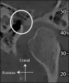
Cone-beam computed tomography image of the right TMJ area (sagittal plane), showing (circle) the petrotympanic fissure morphology type 1 (wide tunnel-shape).
Figure 4
Figure 4
Cone-beam computed tomography image of the right TMJ area (sagittal plane), showing (circle) the petrotympanic fissure morphology type 2 (narrowed tunnel-shaped. Widely open at the entrance of the mandibular fossa and open at the exit in the tympanic cavity, but narrow in the middle aspect).
Figure 5
Figure 5
Cone-beam computed tomography image of the left TMJ area (sagittal plane), showing (circle) the petrotympanic fissure morphology type 3 (very narrow/closed tunnel-shaped. Widely open at the entrance of the mandibular fossa, while the middle region resembles a flat-shaped tunnel structure and narrow (to almost closed) exit in the tympanic cavity).

References

    1. Van Sickels JE, Nishioka GJ, Hegewald MD, Neal GD. Middle ear injury resulting from temporomandibular joint arthroscopy. J Oral Maxillofac Surg. 1987 Nov;45(11):962-5. - DOI - PubMed
    1. Applebaum EL, Berg LF, Kumar A, Mafee MF. Otologic complications following temporomandibular joint arthroscopy. Ann Otol Rhinol Laryngol. 1988 Nov-Dec;97(6 Pt 1):675-9. - DOI - PubMed
    1. Carroll TA, Smith K, Jakubowski J. Extradural haematoma following temporomandibular joint arthrocentesis and lavage. Br J Neurosurg. 2000 Apr;14(2):152-4. - DOI - PubMed
    1. Patel S, Jerjes W, Upile T, Hopper C. TMJ arthroscopy: rare neurological complications associated with breach of the skull base. Br J Oral Maxillofac Surg. 2010 Jun;48(4):e18-20. - DOI - PubMed
    1. Holmlund A, Hellsing G. Arthroscopy of the temporomandibular joint. An autopsy study. Int J Oral Surg. 1985 Apr;14(2):169-75. - DOI - PubMed

LinkOut - more resources