Applications of Computer Technology in Complex Craniofacial Reconstruction
- PMID: 29707444
- PMCID: PMC5908507
- DOI: 10.1097/GOX.0000000000001655
Applications of Computer Technology in Complex Craniofacial Reconstruction
Abstract
Background: To demonstrate our use of advanced 3-dimensional (3D) computer technology in the analysis, virtual surgical planning (VSP), 3D modeling (3DM), and treatment of complex congenital and acquired craniofacial deformities.
Methods: We present a series of craniofacial defects treated at a tertiary craniofacial referral center utilizing state-of-the-art 3D computer technology. All patients treated at our center using computer-assisted VSP, prefabricated custom-designed 3DMs, and/or 3D printed custom implants (3DPCI) in the reconstruction of craniofacial defects were included in this analysis.
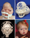
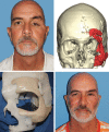
Results: We describe the use of 3D computer technology to precisely analyze, plan, and reconstruct 31 craniofacial deformities/syndromes caused by: Pierre-Robin (7), Treacher Collins (5), Apert's (2), Pfeiffer (2), Crouzon (1) Syndromes, craniosynostosis (6), hemifacial microsomia (2), micrognathia (2), multiple facial clefts (1), and trauma (3). In select cases where the available bone was insufficient for skeletal reconstruction, 3DPCIs were fabricated using 3D printing. We used VSP in 30, 3DMs in all 31, distraction osteogenesis in 16, and 3DPCIs in 13 cases. Utilizing these technologies, the above complex craniofacial defects were corrected without significant complications and with excellent aesthetic results.
Conclusion: Modern 3D technology allows the surgeon to better analyze complex craniofacial deformities, precisely plan surgical correction with computer simulation of results, customize osteotomies, plan distractions, and print 3DPCI, as needed. The use of advanced 3D computer technology can be applied safely and potentially improve aesthetic and functional outcomes after complex craniofacial reconstruction. These techniques warrant further study and may be reproducible in various centers of care.
Conflict of interest statement
Disclosure: The authors have no financial interest to declare in relation to the content of this article. The Article Processing Charge was paid for by the authors.
Figures


References
-
- Hull C; inventor. Apparatus for production of three-dimensional objects by stereolithography. US Patent 4,575,330. March 11, 1986.
-
- Schubert C, van Langeveld MC, Donoso LA. Innovations in 3D printing: a 3D overview from optics to organs. Br J Ophthalmol. 2014;98:159–161.. - PubMed
-
- Doi K. Diagnostic imaging over the last 50 years: research and development in medical imaging science and technology. Phys Med Biol. 2006;51:R5–27.. - PubMed
LinkOut - more resources
Full Text Sources
Other Literature Sources
