Review
. 2018 Mar;6(6):104.
doi: 10.21037/atm.2018.03.24.

Minimally invasive anterior, lateral, and oblique lumbar interbody fusion: a literature review

Affiliations
Review

Minimally invasive anterior, lateral, and oblique lumbar interbody fusion: a literature review

David S Xu et al. Ann Transl Med. 2018 Mar.

Abstract

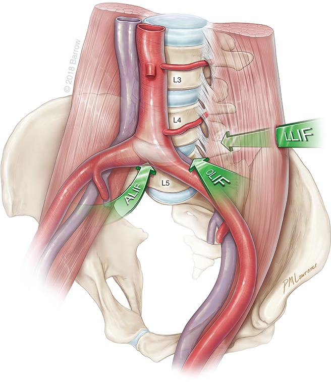
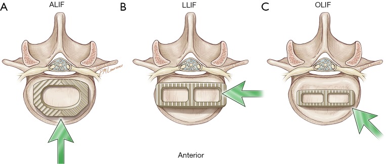
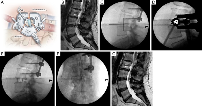
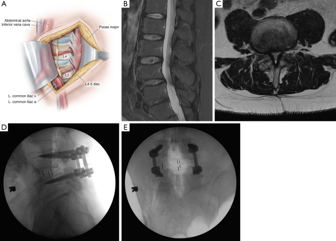
Lumbar interbody fusion involves insertion of a structural graft into an intervertebral disc space to promote bony arthrodesis. It is a well-established surgical strategy for multiple spinal disorders ranging from degenerative conditions to trauma, neoplastic diseases, and deformities requiring correction. Since the inception of lumbar interbody fusion, the most established techniques have been two posterior approaches, the posterior lumbar interbody fusion (PLIF) and the transforaminal lumbar interbody fusion (TLIF). Within the past 15 years, multiple anterolateral approaches to the spine have become widely adopted. These approaches can be performed minimally invasively and spare disruption of the paraspinal muscles and posterior spinal column while enabling wide exposure of the disc space for insertion of interbody grafts much larger than PLIF and TLIF instrumentation. This review highlights three minimally invasive anterolateral approaches: the anterior lumbar interbody fusion (ALIF), the transpsoas lateral lumbar interbody fusion (LLIF), and prepsoas or anterior to the psoas oblique lumbar interbody fusion (OLIF). Relevant topics for discussion and comparison include patient selection, surgical techniques, outcomes, and complications for the three surgical approaches.

Keywords: Anterior lumbar interbody fusion (ALIF); extreme lateral interbody fusion (XLIF); lateral lumbar interbody fusion (LLIF); minimally invasive surgery (MIS); oblique lumbar interbody fusion (OLIF); spinal fusion.


Conflict of interest statement

Conflicts of Interest: Dr. Uribe is a consultant for NuVasive, Inc. The other authors have no conflicts of interest to declare.

Figures

Figure 1
Figure 1
Overview of approaches to the spine. Illustration depicts the three different anterolateral approaches (arrows) to the spine. All three approaches are retroperitoneal. They involve a midline anterior lumbar interbody fusion (ALIF), a lateral lumbar interbody fusion (LLIF), or an oblique lumbar interbody fusion (OLIF). Used with permission from Barrow Neurological Institute, Phoenix, Arizona.
Figure 2
Figure 2
Differences in the footprint of various interbodies. (A) An anterior lumbar interbody fusion (ALIF) fully exposes the anterior span of the disc space and permits insertion of a large interbody graft with a footprint that covers most of the endplate surface area; (B) a lateral lumbar interbody fusion (LLIF) graft is inserted laterally and can be both long and wide, spanning the entire anterior-posterior width of the disc space, including the endplate diaphysis on both sides; (C) an oblique lumbar interbody fusion (OLIF) graft is inserted obliquely in the direction of the contralateral neuroforamen; therefore, its length and width are limited to prevent iatrogenic injury to the nerve root before rotation of the graft into the lateral position. Used with permission from Barrow Neurological Institute, Phoenix, Arizona.
Figure 3
Figure 3
Indirect decompression after lateral lumbar interbody fusion (LLIF). Sagittal T2-weighted magnetic resonance images (A) before and (B) after L1-L3 LLIF for spinal stenosis and coronal deformity in a 69-year-old man with severe back pain and neurogenic claudication. (B) Note expansion of the central canal and visible reduction in nerve root clumping after resolution of the central canal stenosis. Used with permission from Barrow Neurological Institute, Phoenix, Arizona.
Figure 4
Figure 4
Anterior lumbar interbody fusion (ALIF) and reduction of spondylolisthesis. (A) Illustration of a mini-open ALIF approach, in which the peritoneum is reflected laterally to expose the retroperitoneal vessels and spine. At L5-S1, the working corridor is typically between the iliac vessels below their bifurcation. This allows a full anterior-posterior exposure of the disc space in its entirety. At L4–5 and higher lumbar levels, the approach is limited by how much mobilization of the vascular structures can be achieved. a., artery; L. left; v., vein; (B) preoperative sagittal computed tomogram (CT) of a 53-year-old woman with intractable back pain demonstrates a grade I spondylolisthesis at L4–5 that was demonstrated to be mobilized on flexion-extension imaging; (C) postoperative CT of the same patient after a minimally invasive L4–5 ALIF and placement of posterior percutaneous pedicle screws demonstrates full reduction of the spondylolisthesis and clinically significant restoration of disc height. Used with permission from Barrow Neurological Institute, Phoenix, Arizona.
Figure 5
Figure 5
Overview of lateral lumbar interbody fusion (LLIF). (A) Illustration of the LLIF operative field through a minimally invasive retractor allows visualization of most of the lateral length of the disc space, as well as the anterior longitudinal ligament (ALL), which typically is kept intact except when anterior column realignment is necessary. m., muscle; (B) preoperative sagittal T2-weighted magnetic resonance image (MRI) of a 57-year-old woman with a previous L1-L3 fusion and distal adjacent segment instability at L3-4, with resultant severe central canal stenosis causing intractable neurogenic claudication; (C) intraoperative lateral fluoroscopy with a cross hair is used to mark the incision on the patient’s right flank. The center of the incision is approximately one-third of the anterior-posterior (AP) length of the disc space. At L4–5, it is centered on one-half of the AP length; (D) intraoperative lateral fluoroscopy shows the docking location of the minimally invasive lateral retractor; (E) lateral and (F) AP radiographs taken immediately after placement of an LLIF interbody cage demonstrate distraction of the disc space and increased segmental lordosis. A long interbody is placed, spanning the entire wide of the disc space from each diaphysis; (G) postoperative MRI demonstrates restoration of the L3–4-disc height and indirect decompression of the central canal. Used with permission from Barrow Neurological Institute, Phoenix, Arizona.
Figure 6
Figure 6
Overview of an oblique lumbar interbody fusion (OLIF). (A) Illustration of an OLIF exposure, with the medial portion of the psoas major muscle mobilized and retracted posteriorly to expose more of the disc space annulus. The great vessels anterior to the psoas are protected by another retractor. The anterolateral portion of the disc space is then exposed, allowing preparation and insertion of an interbody at an oblique angle that is then rotated to rest laterally; (B) lateral and (C) axial perioperative T2-weighted magnetic resonance images of a 57-year-old man with progressive back pain and neurogenic claudication demonstrate severe L4–5 central canal stenosis and grade I spondylolisthesis; (D) lateral and (E) anterior-posterior radiographs taken during intraoperative placement of the OLIF interbody with percutaneous posterior pedicle screws show good placement of the interbody and reduction of the spondylolisthesis. Note that the width of the interbody does not touch the lateral diaphyses of the disc space. Used with permission from Barrow Neurological Institute, Phoenix, Arizona. a., artery; L. left; m., muscle; v., vein.

References

    1. Briggs H, Milligan PR. Chip fusion of the low back following exploration of the spinal canal. JBJS 1944;26:125-30.
    1. Rao PJ, Maharaj MM, Phan K, et al. Indirect foraminal decompression after anterior lumbar interbody fusion: a prospective radiographic study using a new pedicle-to-pedicle technique. Spine J 2015;15:817-24. 10.1016/j.spinee.2014.12.019 - DOI - PubMed
    1. Oliveira L, Marchi L, Coutinho E, et al. A radiographic assessment of the ability of the extreme lateral interbody fusion procedure to indirectly decompress the neural elements. Spine (Phila Pa 1976) 2010;35:S331-7. 10.1097/BRS.0b013e3182022db0 - DOI - PubMed
    1. Fujibayashi S, Hynes RA, Otsuki B, et al. Effect of indirect neural decompression through oblique lateral interbody fusion for degenerative lumbar disease. Spine 2015;40:E175-E82. 10.1097/BRS.0000000000000703 - DOI - PubMed
    1. Sato J, Ohtori S, Orita S, et al. Radiographic evaluation of indirect decompression of mini-open anterior retroperitoneal lumbar interbody fusion: oblique lateral interbody fusion for degenerated lumbar spondylolisthesis. Eur Spine J 2017;26:671-8. 10.1007/s00586-015-4170-0 - DOI - PubMed