

Melatonin Improves The Developmental Competence of Goat Oocytes
- PMID: 29707934
- PMCID: PMC5936615
- DOI: 10.22074/ijfs.2018.5204
Melatonin Improves The Developmental Competence of Goat Oocytes
Abstract
Background: DNA methylation is one the epigenetic mechanisms, which is critically involved in gene expression. This phenomenon is mediated by DNA methyl-transferases and is affected by environmental stress, including in vitro maturation (IVM) of oocytes. Melatonin, as an antioxidant, may theoretically be involved in epigenetic regulation via reductions of reactive oxygen species. This study was performed to investigate DNA methylation and the possibility of goat oocyte development after treatment with different concentrations of melatonin.
Materials and methods: This experimental study was performed to investigate DNA methylation and the possibility of goat oocyte development after treatment with different concentrations of melatonin. For this purpose, oocytes with granulated cytoplasm were selected and co-cultured with at least two layers of cumulus cells in maturation medium with 10-6 M, 10-9 M, 10-12 M and 0-M (as control group) of melatonin. Nucleus status, glutathione content and developmental competence of the oocytes in each experimental group were assessed. Also, expression of genes associated with DNA methylation, including DNA methyltransferase 1 (DNMT1), DNA methyltransferase 3b (DNMT3b) and DNA methyltransferase 3a (DNMT3a) was evaluated by quantitative real time-polymerase chain reaction (RT-PCR).
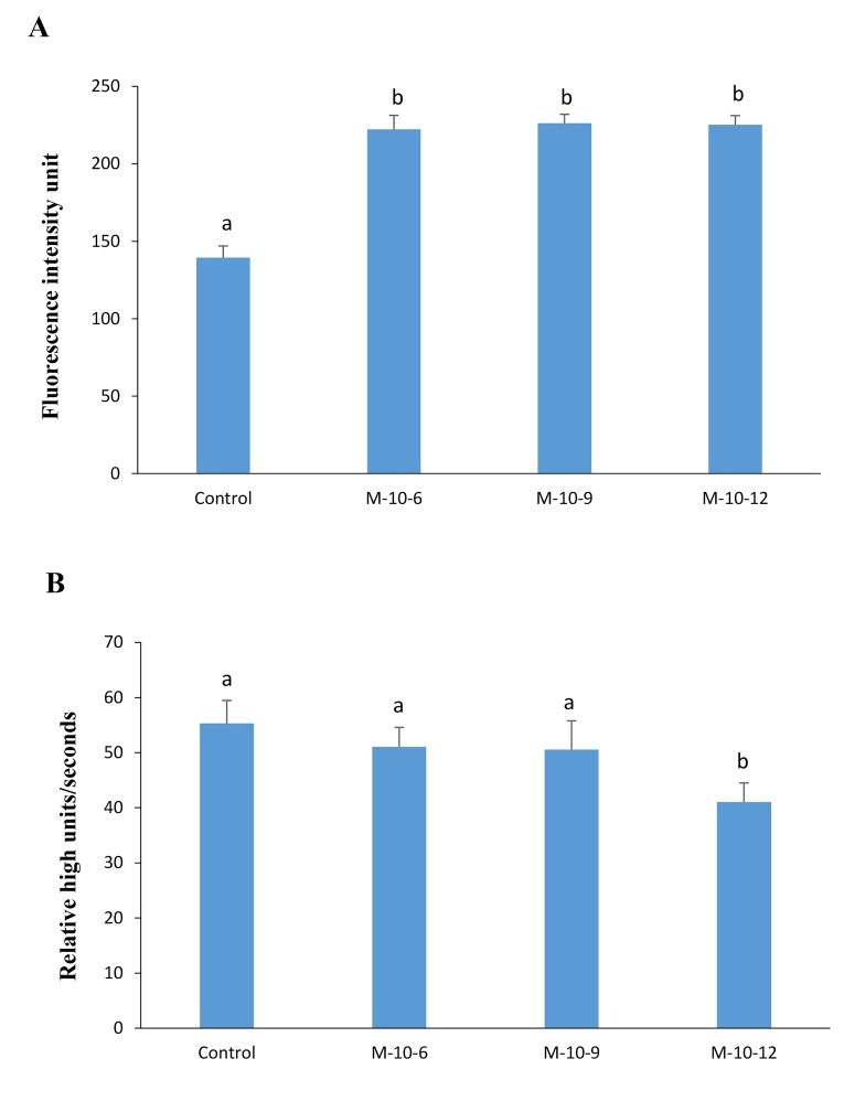
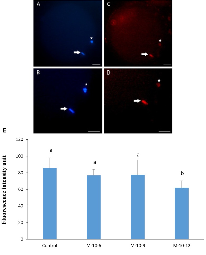
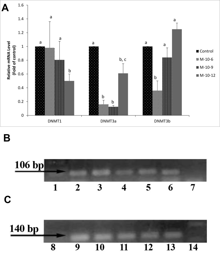
Results: According to our findings, the percentage of oocytes that reached the M-II stage significantly increased in the 10-12 M group (P<0.05). Also, a significant elevation of glutathione content was observed in melatonin-treated oocytes (P<0.05). Analysis of blastocyst formation revealed that developmental competence of the oocytes was higher than the control group (P<0.05). It was observed that melatonin treatment decreased expression levels of DNA methyltransferases (DNMTs) and global DNA methylation (P<0.05). In addition, the expression of melatonin receptor1A (MTNR1A) was detected in both cumulus and oocyte by RT-PCR.
Conclusion: The results suggested that in goat model melatonin affects DNA methylation pattern, leading to an improvement in the developmental competence of the oocytes.
Keywords: Glutathione; Melatonin; Methylation.
Copyright© by Royan Institute. All rights reserved.
Conflict of interest statement
The authors declare no conflicts of interest.
Figures
References
-
- Souza-Fabjan JM, Locatelli Y, Duffard N, Corbin E, Touzé JL, Perreau C, et al. In vitro embryo production in goats: Slaughterhouse and laparoscopic ovum pick up-derived oocytes have different kinetics and requirements regarding maturation media. Theriogenology. 2014;81(8):1021–1031. - PubMed
-
- Russell DF, Baqir S, Bordignon J, Betts DH. The impact of oocyte maturation media on early bovine embryonic development. Mol Reprod Dev. 2006;73(10):1255–1270. - PubMed
-
- Ikeda S, Ichihara-Tanaka K, Azuma T, Muramatsu T, Yamada M. Effects of midkine during in vitro maturation of bovine oocytes on subsequent developmental competence. Biol Reprod. 2000;63(4):1067–1074. - PubMed
-
- De Matos DG, Furnus CC. The importance of having high glutathione (GSH) level after bovine in vitro maturation on embryo development: effect of β-mercaptoethanol, cysteine and cystine. Theriogenology. 2000;53(3):761–771. - PubMed
-
- Abazari-Kia AH, Dehghani-Mohammadabadi M, Mohammadi-Sangcheshmeh A, Zhandi M, Salehi M. Regulation of embryonic development and apoptotic-related gene expression by brain-derived neurotrophic factor in two different culture conditions in ovine. Theriogenology. 2015;84(1):62–69. - PubMed
LinkOut - more resources
Full Text Sources