

Intra-voxel incoherent motion MRI of the living human foetus: technique and test-retest repeatability
- PMID: 29708192
- PMCID: PMC5909359
- DOI: 10.1186/s41747-017-0031-4
Intra-voxel incoherent motion MRI of the living human foetus: technique and test-retest repeatability
Abstract
Background: Our purpose was to test the within-subject (test-retest) reproducibility of the perfusion fraction, diffusion coefficient, and pseudo-diffusion coefficient measurements in various foetus organs and in the placenta based on the intra-voxel incoherent motion (IVIM) principle.
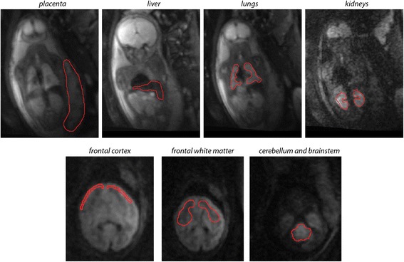
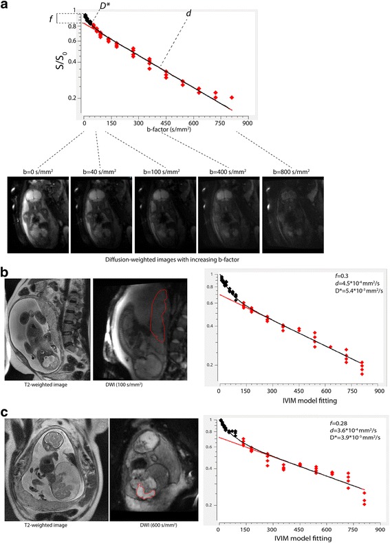
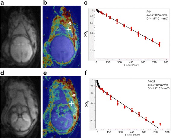
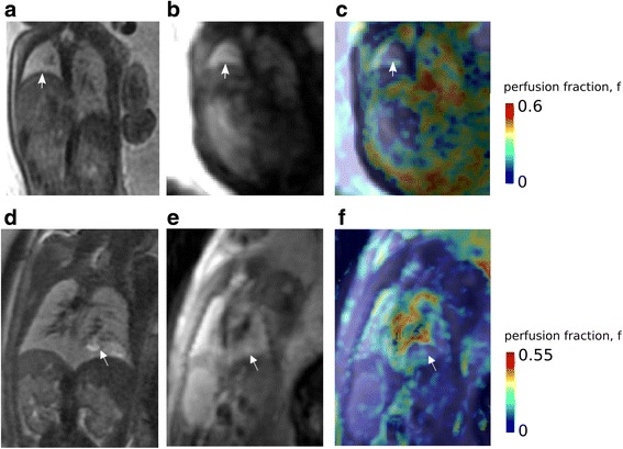
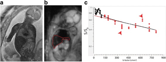
Methods: In utero diffusion-weighted IVIM magnetic resonance imaging (MRI) was performed in 15 pregnant women (pregnancy age 21-36 weeks) on 1.5-T and 3.0-T clinical scanners with b-factors in the range of 0-900 s/mm2 in 16 steps. A bi-exponential model was fitted on the volume-averaged diffusion values. Perfusion fraction (f), diffusion coefficient (d), and pseudo-diffusion coefficient (D*) were calculated. Within-subject reproducibility was evaluated as test-retest variability (VAR %) of the IVIM parameters in the foetal frontal cortex, frontal white matter, cerebellum, lungs, kidneys, liver, and in the placenta.
Results: For the foetal lungs, liver and the placenta, test-retest variability was in the range of 14-20% for f, 12-14% for d, and 17-25% for D*. The diffusion coefficients of the investigated brain regions were moderately to highly reproducible (VAR 5-15%). However, f and D* showed inferior reproducibility compared to corresponding measures for the lungs, liver, and placenta. The IVIM parameters of the foetal kidney were revealed to be highly variable across scans.
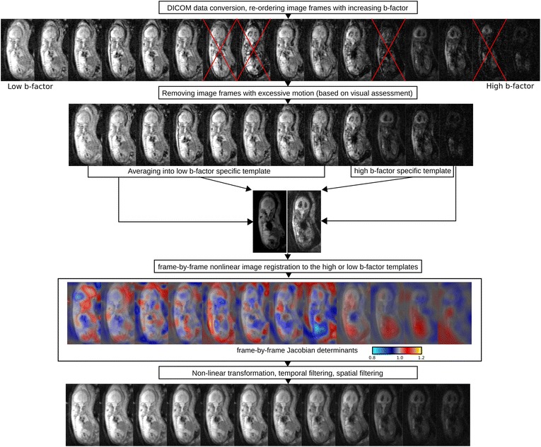
Conclusions: IVIM MRI potentially provides a novel method for examining microvascular perfusion and diffusion in the developing human foetus. However, reproducibility of perfusion and diffusion parameters depends greatly upon data quality, foetal and maternal movements, and foetal-specific image post-processing.
Keywords: Diffusion-weighted imaging (DWI); Foetus; Intra-voxel incoherent motion (IVIM); Magnetic resonance imaging (MRI); Repeatability (reproducibility); Test–retest variability.
Conflict of interest statement
Ethics approval and consent to participateThe mothers gave written informed consent before the MRI examination and the Ethical Commission of Canton Zürich approved the study (EK no. 2017-00167). Not applicable.The authors declare that they have no competing interests.Springer Nature remains neutral with regard to jurisdictional claims in published maps and institutional affiliations.
Figures
Similar articles
-
Microvascular perfusion of the placenta, developing fetal liver, and lungs assessed with intravoxel incoherent motion imaging.J Magn Reson Imaging. 2018 Jul;48(1):214-225. doi: 10.1002/jmri.25933. Epub 2017 Dec 27. J Magn Reson Imaging. 2018. PMID: 29281153
-
Optimization of intra-voxel incoherent motion measurement in diffusion-weighted imaging of breast cancer.J Appl Clin Med Phys. 2017 May;18(3):191-199. doi: 10.1002/acm2.12065. Epub 2017 Mar 27. J Appl Clin Med Phys. 2017. PMID: 28349630 Free PMC article.
-
Comparison of MRI IVIM and MR perfusion imaging in acute ischemic stroke due to large vessel occlusion.Int J Stroke. 2020 Apr;15(3):332-342. doi: 10.1177/1747493019873515. Epub 2019 Sep 3. Int J Stroke. 2020. PMID: 31480940
-
Intravoxel incoherent motion diffusion-weighted MRI of the abdomen: The effect of fitting algorithms on the accuracy and reliability of the parameters.J Magn Reson Imaging. 2017 Jun;45(6):1637-1647. doi: 10.1002/jmri.25535. Epub 2016 Nov 16. J Magn Reson Imaging. 2017. PMID: 27865032
-
Intravoxel incoherent motion diffusion-weighted MR imaging of the liver: effect of triggering methods on regional variability and measurement repeatability of quantitative parameters.Radiology. 2015 Feb;274(2):405-15. doi: 10.1148/radiol.14140759. Epub 2014 Sep 17. Radiology. 2015. PMID: 25232802
Cited by
-
Multi-modal MRI reveals changes in placental function following preterm premature rupture of membranes.Magn Reson Med. 2023 Mar;89(3):1151-1159. doi: 10.1002/mrm.29483. Epub 2022 Oct 18. Magn Reson Med. 2023. PMID: 36255151 Free PMC article.
-
Standard diffusion-weighted, diffusion kurtosis and intravoxel incoherent motion MR imaging of the whole placenta: a pilot study of volumetric analysis.Ann Transl Med. 2022 Mar;10(6):269. doi: 10.21037/atm-22-1037. Ann Transl Med. 2022. PMID: 35434012 Free PMC article.
-
Physiological assessment of the fetal body using MRI: current uses and potential directions.Br J Radiol. 2023 Jul;96(1147):20221024. doi: 10.1259/bjr.20221024. Epub 2023 Jun 13. Br J Radiol. 2023. PMID: 37310734 Free PMC article. Review.
-
The application of in utero magnetic resonance imaging in the study of the metabolic and cardiovascular consequences of the developmental origins of health and disease.J Dev Orig Health Dis. 2021 Apr;12(2):193-202. doi: 10.1017/S2040174420001154. Epub 2020 Dec 14. J Dev Orig Health Dis. 2021. PMID: 33308364 Free PMC article. Review.
-
The haemodynamics of the human placenta in utero.PLoS Biol. 2020 May 28;18(5):e3000676. doi: 10.1371/journal.pbio.3000676. eCollection 2020 May. PLoS Biol. 2020. PMID: 32463837 Free PMC article.
References
LinkOut - more resources
Full Text Sources
Other Literature Sources