. 2018 Jun 8;4(6):918-925.
doi: 10.1021/acsinfecdis.8b00044. Epub 2018 May 7.

Compartment-Specific Labeling of Bacterial Periplasmic Proteins by Peroxidase-Mediated Biotinylation

Compartment-Specific Labeling of Bacterial Periplasmic Proteins by Peroxidase-Mediated Biotinylation

Uday S Ganapathy et al. ACS Infect Dis. .

Erratum in

Abstract

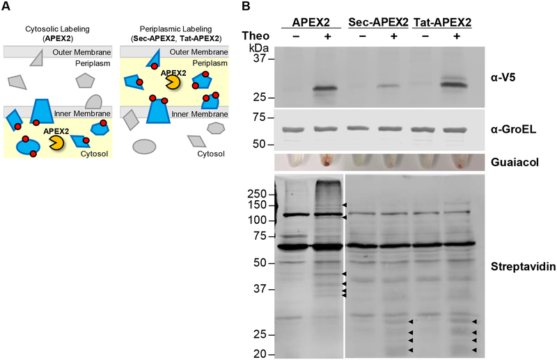
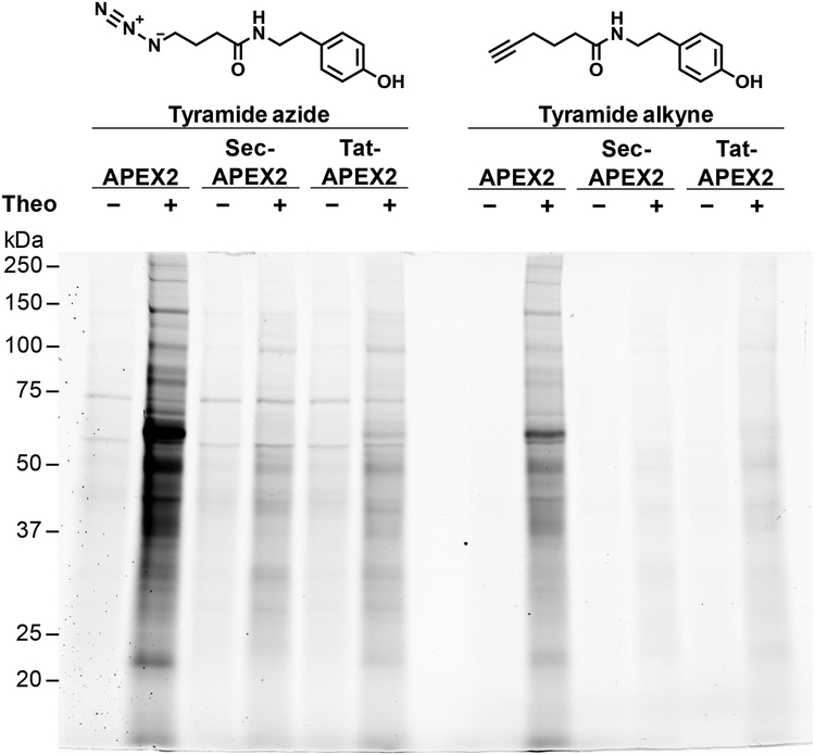
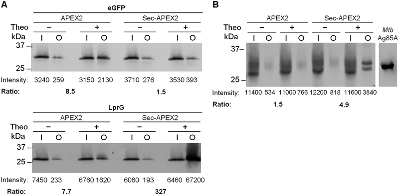
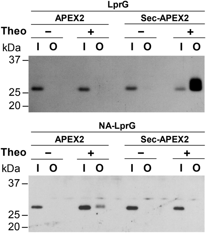
The study of the bacterial periplasm requires techniques with sufficient spatial resolution and sensitivity to resolve the components and processes within this subcellular compartment. Peroxidase-mediated biotinylation has enabled targeted labeling of proteins within subcellular compartments of mammalian cells. We investigated whether this methodology could be applied to the bacterial periplasm. In this study, we demonstrated that peroxidase-mediated biotinylation can be performed in mycobacteria and Escherichia coli. To eliminate detection artifacts from natively biotinylated mycobacterial proteins, we validated two alternative labeling substrates, tyramide azide and tyramide alkyne, which enable biotin-independent detection of labeled proteins. We also targeted peroxidase expression to the periplasm, resulting in compartment-specific labeling of periplasmic versus cytoplasmic proteins in mycobacteria. Finally, we showed that this method can be used to validate protein relocalization to the cytoplasm upon removal of a secretion signal. This novel application of peroxidase-mediated protein labeling will advance efforts to characterize the role of the periplasm in bacterial physiology and pathogenesis.

Keywords: APEX; CuAAC click chemistry; bacteria; mycobacteria; periplasm; peroxidase-mediated biotinylation.


Figures

Figure 1.
Figure 1.. Cytoplasmic and secreted APEX2 have distinct protein biotinylation profiles in Msm.
(A) To localize APEX2 to the periplasmic space, we generated Sec-APEX2 and Tat-APEX2, in which a Sec or Tat secretion pathway signal is fused to APEX2. Red circles denote labeling substrate that reacts covalently with proteins in an APEX2 activity-dependent manner and subsequently serves as an enrichment or detection tag. (B) Msm expressing V5-tagged APEX2 (28 kDa), Sec-APEX2 (31 kDa) or Tat-APEX2 (33 kDa) was grown without or with theophylline and subjected to the labeling protocol with biotin-phenol. Anti-V5 immunoblot analysis of the lysates established expression of APEX2, Sec-APEX2 and Tat-APEX2. Anti-GroEL was used to confirm equal loading. Peroxidase activity was assayed in whole cells using guaiacol. Streptavidin blot analysis was used to detect protein biotinylation. Arrowheads indicate examples of APEX2 expression-dependent bands. Data are representative of >3 biological replicates.
Figure 2.
Figure 2.. Alternative labeling substrates reduce detection artifacts.
Msm expressing APEX2, Sec-APEX2 or Tat-APEX2 was grown without or with theophylline and subjected to the labeling protocol with either tyramide azide or tyramide alkyne. Lysates underwent a CuAAC reaction with alkyne or azide-conjugated fluorescein. In-gel fluorescence detection of the clicked lysates was used to assess protein labeling. Equal protein loading was confirmed by subsequent Coomassie staining (Figure S5). Data are representative of 3 biological replicates under the specified conditions.
Figure 3.
Figure 3.. Protein biotinylation by APEX2 and Sec-APEX2 is compartment-specific.
(A) Msm co-expressing APEX2 or Sec-APEX2 from a single integrated chromosomal copy and eGFP-3XFLAG (31 kDa) or LprG-3XFLAG (27 kDa) from a multi-copy episomal plasmid were grown without or with theophylline and subjected to the labeling protocol with biotin-phenol. Biotinylated proteins were enriched by avidin affinity purification. Anti-FLAG immunoblot analysis with fluorescence detection was used to quantify expression and biotinylation. The fold increase in biotinylation upon induction of APEX2 or Sec-APEX2 (“Ratio”) was calculated by taking the ratio of the + / − theophylline output signals after normalizing to the corresponding inputs. (B) Msm expressing APEX2 or Sec-APEX2 from a multi-copy episomal plasmid were treated as in (A) except that immunoblot analysis was performed with antibody against Mtb Ag85 complex. Purified Mtb Ag85A (34 kDa) was included as a positive control for the antibody. All data are from the same blot; intervening lanes were removed for clarity. All data are representative of ≥ 2 biological replicates. Lane labels I and O indicate input and output for the avidin enrichment.
Figure 4.
Figure 4.. Truncating the predicted secretion signal relocalizes LprG to the cytoplasm.
Msm co-expressing APEX2 or Sec-APEX2 from a multi-copy episomal plasmid and LprG-3XFLAG (27 kDa) or NA-LprG-3XFLAG (24 kDa) from a single integrated chromosomal copy were grown without or with theophylline and subjected to the labeling protocol with biotin-phenol. Biotinylated proteins were enriched by avidin affinity purification. Anti-FLAG immunoblot analysis with chemiluminescence detection of the input and output fractions was used to assess expression and biotinylation of LprG-3XFLAG and NA-LprG-3XFLAG. Data are representative of 3 biological replicates. Lane labels I and O indicate input and output for the avidin enrichment.

References

    1. Weiner JH; Li L, Proteome of the Escherichia coli envelope and technological challenges in membrane proteome analysis. Biochim Biophys Acta 2008, 1778 (9), 1698–1713. DOI: 10.1016/j.bbamem.2007.07.020. - DOI - PubMed
    1. Belardinelli JM; Larrouy-Maumus G; Jones V; Sorio de Carvalho LP; McNeil MR; Jackson M, Biosynthesis and translocation of unsulfated acyltrehaloses in Mycobacterium tuberculosis. J. Biol. Chem 2014, 289 (40), 27952–27965. DOI: 10.1074/jbc.M114.581199. - DOI - PMC - PubMed
    1. Seeliger JC; Holsclaw CM; Schelle MW; Botyanszki Z; Gilmore SA; Tully SE; Niederweis M; Cravatt BF; Leary JA; Bertozzi CR, Elucidation and chemical modulation of sulfolipid-1 biosynthesis in Mycobacterium tuberculosis. J. Biol. Chem 2012, 287 (11), 7990–8000. DOI: 10.1074/jbc.M111.315473. - DOI - PMC - PubMed
    1. Touchette MH; Holsclaw CM; Previti ML; Solomon VC; Leary JA; Bertozzi CR; Seeliger JC, The rv1184c locus encodes Chp2, an acyltransferase in Mycobacterium tuberculosis polyacyltrehalose lipid biosynthesis. J. Bacteriol 2015, 197 (1), 201–210. DOI: 10.1128/JB.02015-14. - DOI - PMC - PubMed
    1. McCann JR; McDonough JA; Sullivan JT; Feltcher ME; Braunstein M, Genome-Wide Identification of Mycobacterium tuberculosis Exported Proteins with Roles in Intracellular Growth. J. Bacteriol 2011, 193 (4), 854–861. DOI: 10.1128/jb.01271-10. - DOI - PMC - PubMed

Publication types

MeSH terms