Case Reports
. 2018 Apr 30;12(4):e0006429.
doi: 10.1371/journal.pntd.0006429. eCollection 2018 Apr.

Microdeletion on chromosome 8p23.1 in a familial form of severe Buruli ulcer

Affiliations
Case Reports

Microdeletion on chromosome 8p23.1 in a familial form of severe Buruli ulcer

Quentin B Vincent et al. PLoS Negl Trop Dis. .

Abstract

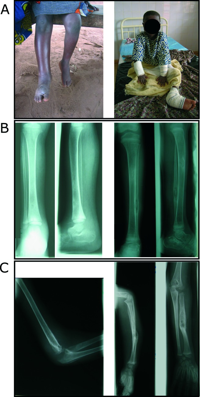
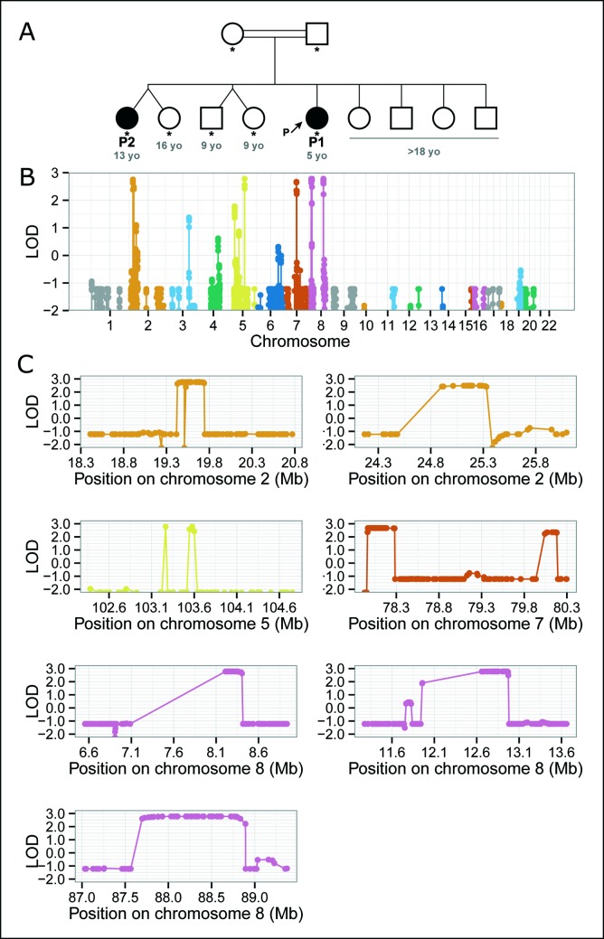
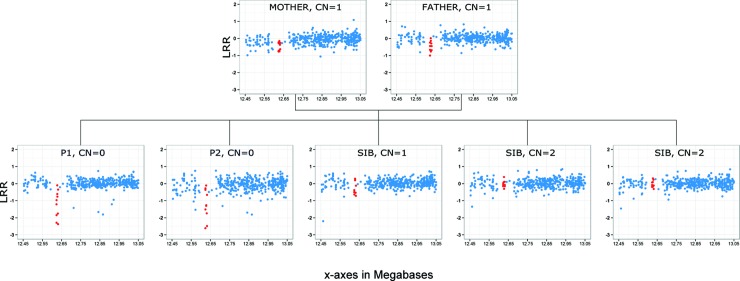
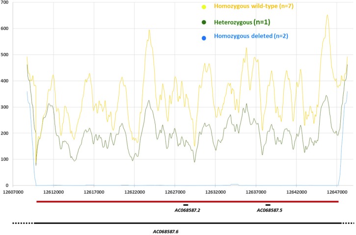
Buruli ulcer (BU), the third most frequent mycobacteriosis worldwide, is a neglected tropical disease caused by Mycobacterium ulcerans. We report the clinical description and extensive genetic analysis of a consanguineous family from Benin comprising two cases of unusually severe non-ulcerative BU. The index case was the most severe of over 2,000 BU cases treated at the Centre de Dépistage et de Traitement de la Lèpre et de l'Ulcère de Buruli, Pobe, Benin, since its opening in 2003. The infection spread to all limbs with PCR-confirmed skin, bone and joint infections. Genome-wide linkage analysis of seven family members was performed and whole-exome sequencing of both patients was obtained. A 37 kilobases homozygous deletion confirmed by targeted resequencing and located within a linkage region on chromosome 8 was identified in both patients but was absent from unaffected siblings. We further assessed the presence of this deletion on genotyping data from 803 independent local individuals (402 BU cases and 401 BU-free controls). Two BU cases were predicted to be homozygous carriers while none was identified in the control group. The deleted region is located close to a cluster of beta-defensin coding genes and contains a long non-coding (linc) RNA gene previously shown to display highest expression values in the skin. This first report of a microdeletion co-segregating with severe BU in a large family supports the view of a key role of human genetics in the natural history of the disease.


Conflict of interest statement

The authors have declared that no competing interests exist.

Figures

Fig 1
Fig 1. Clinical course of unusually severe BU in the index patient P1.
All four limbs were affected by edematous BU lesions, with osteomyelitis involving ten bones and septic arthritis involving two joints. (A) Severe edema of the right leg and foot and edema of the left foot (left panel) leading to amputation of the lower right leg (right panel). (B) Mediodiaphyseal lacuna of the lower third of the right tibia and major soft tissue enlargement (left panel). Hypercondensation of the proximal third of the left tibia and lacuna of the lower third of the fibular diaphysis with central sequestrum (right panel). (C) Intra-articular effusion of the right elbow associated to osteolysis and periostal reaction of the humerus, the olecranon and the radial head (left panel). Forearm diaphyseal lacuna of the left radius with sequestrum and partly consolidated fracture of the left radius (right panel).
Fig 2
Fig 2. BU of unusual severity segregates in a consanguineous family from Benin and maps to chromosomes 2, 5, 7 and 8.
(A) Pedigree tree. Genotyped individuals are indicated by stars with age in years (blue numbers) given at the time of diagnosis for the two patients and at the time of the study for unaffected individuals. Two of the nine siblings were affected and the parents were consanguineous. We therefore hypothesized a recessive mode of inheritance, and carried out linkage analysis by homozygosity mapping. Note that P2’s twin is not an identical twin. (B) Genome-wide linkage analysis by homozygosity mapping. This statistical approach aims to identify genomic regions homozygous and identical-by-descent in affected individuals but not homozygous in unaffected individuals. The evidence for linkage is based on LOD scores (y axis). (C) Linkage regions on chromosomes 2 (two regions: 2.1, 2.2), 5, 7 (7.1, 7.2), and 8 (8.1, 8.2, 8.3). Homozygosity mapping identifies 8 linkage regions spanning hundreds of kbs to Mbs very likely to contain the causal genetic lesion but does not identify the genetic lesion itself. For the exact coordinates of the linkage regions, see S1 Table.
Fig 3
Fig 3. Genome-wide CNV analysis identifies a homozygous deletion in the second linkage region on chromosome 8.
CNV identification is based principally on quantitative analysis of the intensity of the hybridization signal of nucleotide probes (the logR ratio, LRR) throughout the genome, making it possible to infer that the copy number (CN) is normal, increased or decreased in the patient’s genome, at the location of the probe. Each blue dot represents one probe in this region of chromosome 8. Red dots represent the CNV segment identified in this family by PennCNV (from probe CN_1273661 to probe CN_1273690, at position 12,616,022 to 12,624,550 in GRCh38 coordinates).
Fig 4
Fig 4. Distribution of the mean coverage per base in the 8q32 deletion region according to the deletion status.
Zoom out of the deletion region (Chr8:12,607,000–12,647,000). Positions in base pair (bp) are given on the x-axis. Y-axis displays the mean coverage per base as estimated through 1kb sliding windows (i.e. value at position X is the mean over X +/- 500 bp; note for X<1kb mean is over X + 1kb). Custom tracks shows the delineated deletion (Chr8:12,609,841–12,647,341—horizontal red bar), and the genes reported from the Vega database as implemented in the Ensembl genome browser (horizontal black lines): AC068587.6 (Ensembl gene ID: ENSG00000283674); AC068587. 2 (ENSG00000244289); AC068587.5 (ENSG00000255253).

References

    1. Sizaire V, Nackers F, Comte E, Portaels F. Mycobacterium ulcerans infection: control, diagnosis, and treatment. Lancet Infect Dis. 2006;6(5):288–96. Epub 2006/04/25. S1473-3099(06)70464-9 [pii] doi: 10.1016/S1473-3099(06)70464-9 . - DOI - PubMed
    1. World Health Organization. Buruli ulcer disease factsheet 2017. Available from: http://www.who.int/mediacentre/factsheets/fs199/en/index.html
    1. Eddyani M, Sopoh GE, Ayelo G, Brun LVC, Roux JJ, Barogui Y, et al. Diagnostic Accuracy of Clinical and Microbiological Signs in Patients with Skin Lesions Resembling Buruli Ulcer in an Endemic Region. Clin Infect Dis. 2018. doi: 10.1093/cid/ciy197 . - DOI - PMC - PubMed
    1. Vincent QB, Ardant MF, Adeye A, Goundote A, Saint-Andre JP, Cottin J, et al. Clinical epidemiology of laboratory-confirmed Buruli ulcer in Benin: a cohort study. Lancet Glob Health. 2014;2(7):e422–30. Epub 2014/08/12. S2214-109X(14)70223-2 [pii] doi: 10.1016/S2214-109X(14)70223-2 . - DOI - PubMed
    1. Vincent QB, Ardant MF, Marsollier L, Chauty A, Alcais A. HIV infection and Buruli ulcer in Africa. Lancet Infect Dis. 2014;14(9):796–7. Epub 2014/08/29. S1473-3099(14)70882-5 [pii] doi: 10.1016/S1473-3099(14)70882-5 . - DOI - PubMed

Publication types