. 2018 Apr 30;13(4):e0193907.
doi: 10.1371/journal.pone.0193907. eCollection 2018.

Development and characterization of carboxy-terminus specific monoclonal antibodies for understanding MUC16 cleavage in human ovarian cancer

Affiliations

Development and characterization of carboxy-terminus specific monoclonal antibodies for understanding MUC16 cleavage in human ovarian cancer

Abhijit Aithal et al. PLoS One. .

Abstract

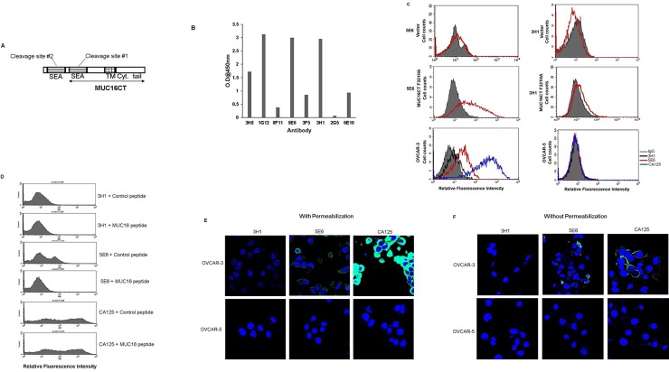
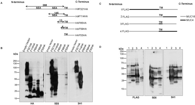
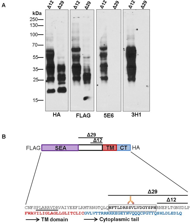
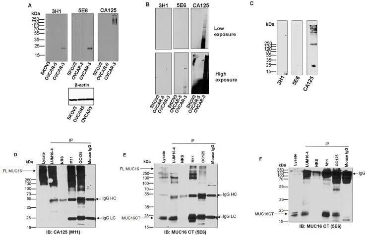
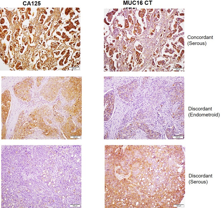
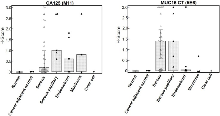
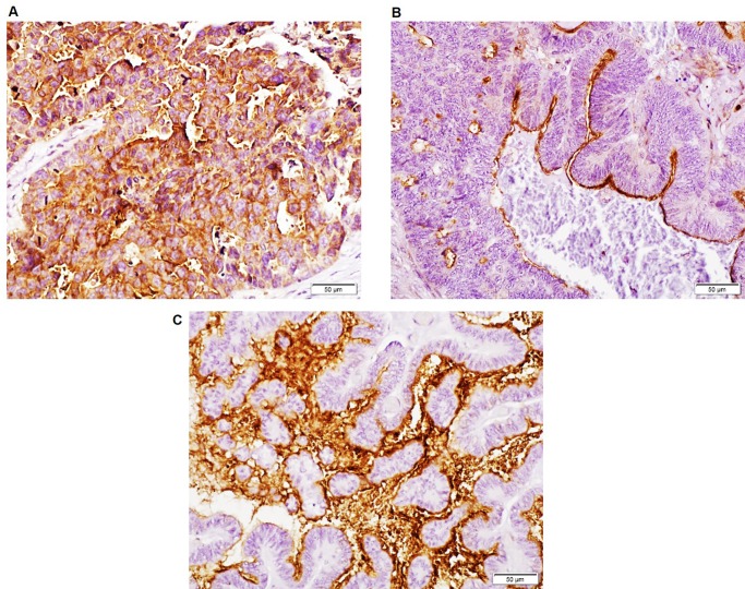
MUC16 is overexpressed in ovarian cancer and plays important roles in invasion and metastasis. Previously described monoclonal antibodies against cell surface expressed MUC16 recognize the N-terminal tandemly repeated epitopes present in cancer antigen 125 (CA125). MUC16 is cleaved at a specific location, thus, releasing CA125 into the extracellular space. Recent reports have indicated that the retained carboxy-terminal (CT) fragment of MUC16 might play an important role in tumorigenicity in diverse types of cancers. However, limited data is available on the fate and existence of CT fragment on the surface of the cancer cell. Herein, we characterize two monoclonal antibodies (mAbs) showing specificity to the retained juxtamembrane region of MUC16. For the first time, we demonstrate that MUC16 is cleaved in ovarian cancer cells (NIH:OVCAR-3 [OVCAR-3]) and that the cleaved MUC16 subunits remain associated with each other. Immunohistochemical analyses on different grades of ovarian tumor tissues indicated differential reactivity of CA125 and MUC16 CT mAbs. The CA125 (M11) mAb detected 32/40 (80%), while the CT mAb (5E6) detected 33/40 (82.5%) of total ovarian cancer cases. For serous and serous papillary cases, the CA125 (M11) mAb stained 27/31 cases (87%), while CT mAb (5E6) stained 29/31 cases (93.5%). The CT mAb(s) accurately predict expression of MUC16 since their epitopes are not tandemly repeated and their reactivity may not be dependent on O-linked glycosylation. These antibodies can serve as valuable reagents for understanding MUC16 cleavage and may also serve as potential therapeutic agents for treatment of ovarian cancer.


Conflict of interest statement

Competing Interests: Dr. Wade Junker is a paid employee of Sanguine Diagnostics and Therapeutics Inc. and supported by the indicated NIH grants awarded to the company. The specific roles of these authors are articulated in the 'author contributions' section. This does not alter our adherence to PLOS ONE policies on sharing data and materials.

Figures

Fig 1
Fig 1. Generation and characterization of monoclonal antibodies (mAbs) to MUC16 C-terminal (CT) domain.
(A) Structure of MUC16 CT domain indicating the two membrane-proximal cleavage sites. The fragment used for generation of hybridomas is indicated by a line with double arrow heads. This fragment incorporates the last putative cleavage site. Other important domains are indicated: TM- Transmembrane domain; Cyt. Tail- Cytoplasmic tail. (B) Binding of selected anti-MUC16 CT monoclonal antibodies with purified MUC16 CT protein using an indirect ELISA. MUC16 CT protein was coated in ELISA wells and hybridoma supernatants of the indicated antibodies were added. Antibody binding was detected using a secondary antibody labeled with horseradish peroxidase and TMB substrate. (C) Flow cytometry analysis showing relative binding of anti-MUC16 CT mAbs (5E6 and 3H1) to MIA PaCa-2 cells transfected either with control vector or MUC16 CT FL 321 construct (last 321 amino acids of MUC16). Binding was also analyzed on OVCAR-3 (MUC16HIGH) and OVCAR-5 (MUC16LOW) cells. Anti- tandem repeat mAb M11 served as a positive control. Cells were stained with the indicated antibodies and the signal was detected using Alexa-Fluor 488 anti-mouse IgG secondary antibody. A mouse IgG1 antibody served as the irrelevant isotype control and is indicated by the gray shaded curve. (D) Flow cytometry analysis of OVCAR-3 cells using mAbs pre-incubated with either MUC16 peptide or irrelevant control peptide. (E and F) OVCAR-3 and OVCAR-5 cells were seeded on coverslips, fixed with 4% Paraformaldehyde in PBS and were either permeabilized with 0.1% Triton X-100 in PBS (E) or not permeabilized (F) and incubated with 10 μg/ml of indicated mAbs. Signal was detected using Alexa Fluor 488 conjugated secondary antibody. Coverslips were placed on glass slides containing a drop of anti-fade Vectashield mounting medium and observed under a ZEISS confocal laser scanning microscope (magnification, 630X).
Fig 2
Fig 2. Partial epitope mapping using MUC16 CT constructs transfected into HEK293T cells.
(A) Schematic representation of different lengths of MUC16 CT fragments with C-terminal HA-tag cloned into the p3X-FLAG-CMV9 vector (Empty vector) with a preprotrypsin leader peptide (LP). The predicted cleavage sites in the last (site #1, PLARRVDR) and penultimate (site #2, DSVLV) SEA domains and the transmembrane (TM) domain are indicated. (B) Partial epitope mapping using various constructs of MUC16 CT (given in (A)) transfected into HEK293T cells was performed. Lysates from transfected cells were immunoblotted with the indicated antibodies. (C) The Flag tagged F114HA MUC16 CT construct was domain swapped with the various domains of MUC4 as indicated. (D) The constructs described in (C) were transfected into HEK293T cells. Lysates from these cells were immunoblotted with the respective antibodies.
Fig 3
Fig 3. Narrowing down the epitope recognized by MUC16 CT mAbs.
(A) The last 12 and 29 amino acids from TM domain of MUC16 were deleted in the F114HA construct, tagged with FLAG at N-terminus and HA at C-terminus, to generate 2 deletion constructs (Δ12 and Δ29). The Δ12 and Δ29 constructs were transfected into HEK293T cells and immunoblotted with the indicated antibodies. (B) Schematic representation of MUC16 C-terminal region indicating various domains, cleavage site and approximate location of the putative epitope recognized by mAbs 5E6and 3H1. Key: Purple—represents the last SEA domain. Red portion—indicates the start of the transmembrane domain. Boxed region—indicates the putative epitope. Underlined region—represents the predicted putative cleavage site.
Fig 4
Fig 4. MUC16 is cleaved and a fraction of cleaved fragments remain non-covalently associated in the cells.
Lysates of ovarian cancer cell lines (SKOV3, OVCAR-5 and OVCAR-3) were resolved on 12% SDS-PAGE (A) or 0.1% SDS-2% agarose gels (B), transferred to PVDF membranes and probed with the indicated antibodies. (C) OVCAR-3 culture supernatant was resolved on a gradient (4–12%) SDS-PAGE, transferred to PVDF membranes and probed with the indicated antibodies. (D, E and F) OVCAR-3 cells were lysed and precipitated with MUC16 CT antiserum (LUM16-4) or CA125 mAbs (M11 and OC125) as described in the Materials and Methods section. The precipitates were resolved by SDS-PAGE under reducing (D and E) and non-reducing conditions (F) followed by transfer to PVDF membrane and probed with the indicated antibodies. Mouse IgG1 and non-specific rabbit serum (NRS) were used as negative controls. Full length MUC16 (FL MUC16) was detected as a high molecular weight smear recognized by both CA125 and 5E6 mAbs. MAb 5E6 recognized the cleaved C-terminal fragment of MUC16 (MUC16 CT). Intact non-reduced immunoglobulin (IgG) and reduced heavy and light chains (IgG HC and IgG LC respectively) are indicated.
Fig 5
Fig 5. Comparative analysis of CA125 and MUC16 CT staining in human ovarian cancer cases by IHC.
Duplicate ovarian cancer tissue microarrays (OV1004, BIOMAX) were processed for IHC staining as described in the Materials and Methods section and with primary antibodies CA125 (M11) and MUC16 CT (5E6). Representative cases of concordant and discordant staining are indicated. The top and bottom panels represent serous subtypes which exhibited intense staining with both CA125 and MUC16 CT mAbs. The middle panel represents the endometroid subtype that exhibited positive reactivity with CA125 but demonstrated comparatively lower staining with MUC16 CT mAb.
Fig 6
Fig 6. Box plot comparing CA125 and MUC16 CT staining across various histologic types of ovarian cancer.
MUC16 immunoreactivity was higher in the serous and serous papillary adenocarcinoma tissues as compared to other types. The CA125 mAb also exhibited reactivity with endometroid and mucinous types while CT mAb failed to detect these types efficiently. No staining is observed in normal or cancer adjacent normal ovarian tissue with either CA125 or MUC16 CT mAbs.
Fig 7
Fig 7. MUC16 CT mAb 5E6 exhibits heterogeneous staining on human ovarian cancer tissues.
Sections of ovarian cancer tissues indicating strong membranous and cytoplasmic staining in tumor cells (A), focal apical membranous staining on tumor cells (B), and strong intra-luminal and membranous staining (C) of MUC16 CT. In all cases, the surrounding stroma was negative for MUC16 CT expression. Original magnification 200х.

References

    1. Bast RC Jr., Feeney M, Lazarus H, Nadler LM, Colvin RB, Knapp RC. Reactivity of a monoclonal antibody with human ovarian carcinoma. The Journal of clinical investigation. 1981;68(5):1331–7. doi: 10.1172/JCI110380 - DOI - PMC - PubMed
    1. Yin BW, Dnistrian A, Lloyd KO. Ovarian cancer antigen CA125 is encoded by the MUC16 mucin gene. International journal of cancer. 2002;98(5):737–40. - PubMed
    1. Yin BW, Lloyd KO. Molecular cloning of the CA125 ovarian cancer antigen: identification as a new mucin, MUC16. The Journal of biological chemistry. 2001;276(29):27371–5. doi: 10.1074/jbc.M103554200 - DOI - PubMed
    1. O'Brien TJ, Beard JB, Underwood LJ, Dennis RA, Santin AD, York L. The CA 125 gene: an extracellular superstructure dominated by repeat sequences. Tumour biology: the journal of the International Society for Oncodevelopmental Biology and Medicine. 2001;22(6):348–66. - PubMed
    1. Das S, Rachagani S, Torres-Gonzalez MP, Lakshmanan I, Majhi PD, Smith LM, et al. Carboxyl-terminal domain of MUC16 imparts tumorigenic and metastatic functions through nuclear translocation of JAK2 to pancreatic cancer cells. Oncotarget. 2015;6(8):5772–87. doi: 10.18632/oncotarget.3308 - DOI - PMC - PubMed

Publication types