

Genetic profiling of young and aged endothelial progenitor cells in hypoxia
- PMID: 29708992
- PMCID: PMC5927426
- DOI: 10.1371/journal.pone.0196572
Genetic profiling of young and aged endothelial progenitor cells in hypoxia
Abstract
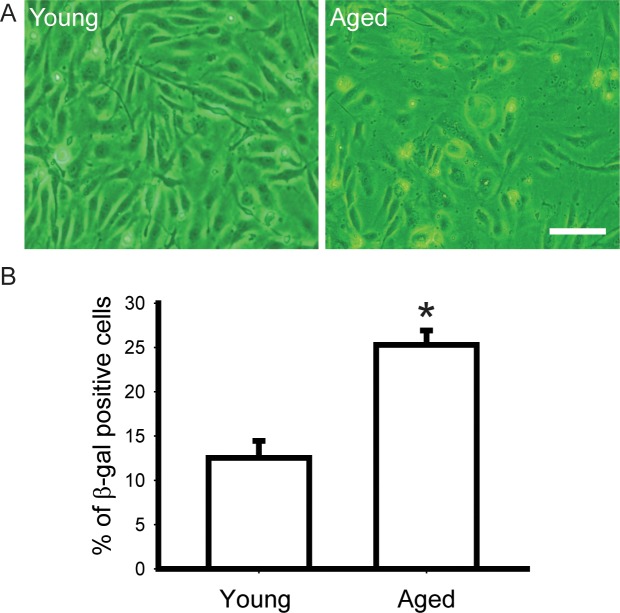
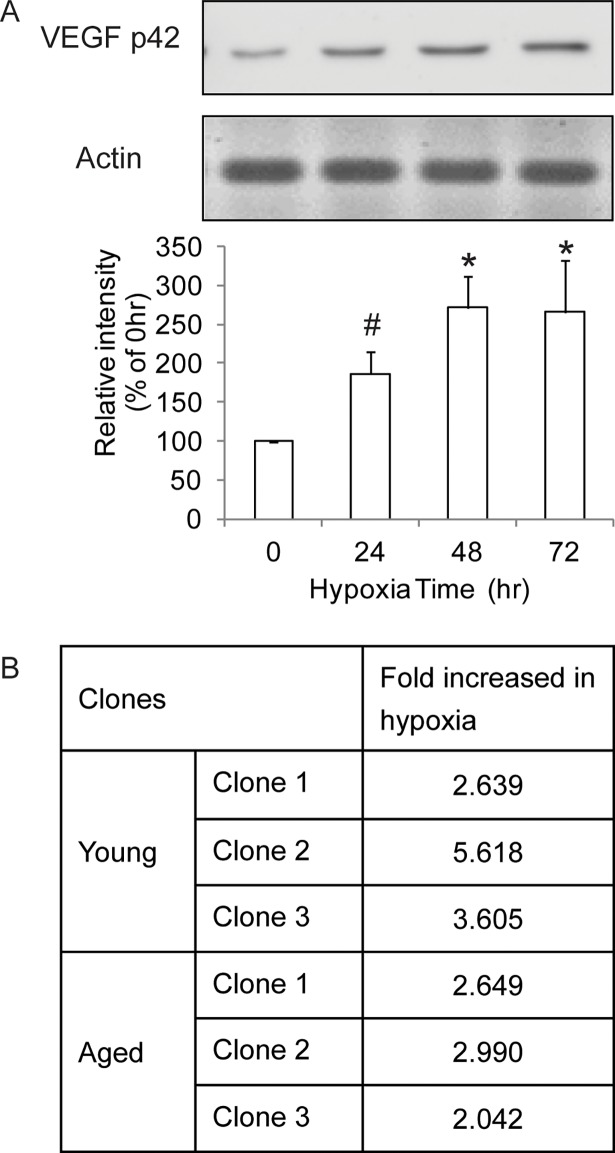
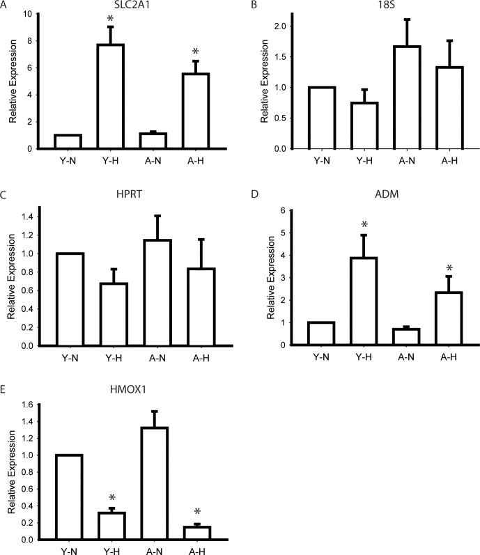
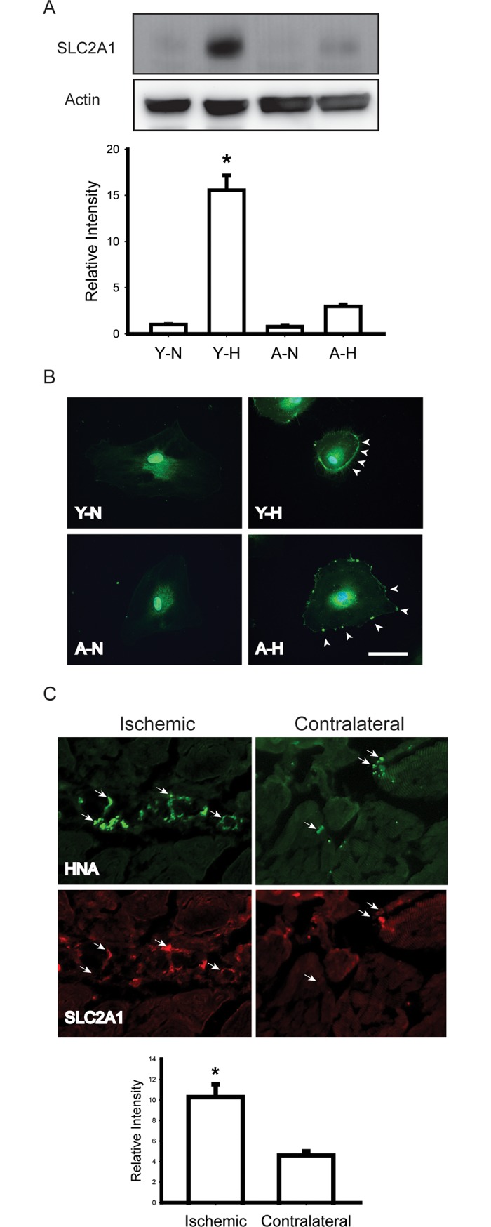
Age is a major risk factor for diseases caused by ischemic hypoxia, such as stroke and coronary artery disease. Endothelial progenitor cells (EPCs) are the major cells respond to ischemic hypoxia through angiogenesis and vascular remodeling. However, the effect of aging on EPCs and their responses to hypoxia are not well understood. CD34+ EPCs were isolated from healthy volunteers and aged by replicative senescence, which was to passage cells until their doubling time was twice as long as the original cells. Young and aged CD34+ EPCs were exposed to a hypoxic environment (1% oxygen for 48hrs) and their gene expression profiles were evaluated using gene expression array. Gene array results were confirmed using quantitative polymerase chain reaction, Western blotting, and BALB/c female athymic nude mice hindlimb ischemia model. We identified 115 differentially expressed genes in young CD34+ EPCs, 54 differentially expressed genes in aged CD34+ EPCs, and 25 common genes between normoxia and hypoxia groups. Among them, the expression of solute carrier family 2 (facilitated glucose transporter), member 1 (SLC2A1) increased the most by hypoxia in young cells. Gene set enrichment analysis indicated the pathways affected by aging and hypoxia most, including genes "response to oxygen levels" in young EPCs and genes involved "chondroitin sulfate metabolic process" in aged cells. Our study results indicate the key factors that contribute to the effects of aging on response to hypoxia in CD34+ EPCs. With the potential applications of EPCs in cardiovascular and other diseases, our study also provides insight on the impact of ex vivo expansion might have on EPCs.
Conflict of interest statement
Figures
References
-
- Charles ST, Carstensen LL (2010) Social and emotional aging. Annu Rev Psychol 61: 383–409. doi: 10.1146/annurev.psych.093008.100448 - DOI - PMC - PubMed
-
- Kelly TN, Bazzano LA, Fonseca VA, Thethi TK, Reynolds K, He J (2009) Systematic review: glucose control and cardiovascular disease in type 2 diabetes. Ann Intern Med 151: 394–403. - PubMed
-
- Kim SY, Guevara JP, Kim KM, Choi HK, Heitjan DF, Albert DA (2009) Hyperuricemia and risk of stroke: a systematic review and meta-analysis. Arthritis Rheum 61: 885–892. doi: 10.1002/art.24612 - DOI - PMC - PubMed
-
- Kuper H, Nicholson A, Kivimaki M, Aitsi-Selmi A, Cavalleri G, Deanfield JE, et al. (2009) Evaluating the causal relevance of diverse risk markers: horizontal systematic review. BMJ 339: b4265 doi: 10.1136/bmj.b4265 - DOI - PMC - PubMed
-
- Milicevic Z, Raz I, Beattie SD, Campaigne BN, Sarwat S, Gromniak E, et al. (2008) Natural history of cardiovascular disease in patients with diabetes: role of hyperglycemia. Diabetes Care 31 Suppl 2: S155–160. - PubMed
Publication types
MeSH terms
Substances
LinkOut - more resources
Full Text Sources
Other Literature Sources
Miscellaneous
