MCU-knockdown attenuates high glucose-induced inflammation through regulating MAPKs/NF-κB pathways and ROS production in HepG2 cells
- PMID: 29709004
- PMCID: PMC5927441
- DOI: 10.1371/journal.pone.0196580
MCU-knockdown attenuates high glucose-induced inflammation through regulating MAPKs/NF-κB pathways and ROS production in HepG2 cells
Abstract
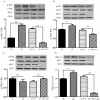
Mitochondrial Ca2+ is a key regulator of organelle physiology and the excessive increase in mitochondrial calcium is associated with the oxidative stress. In the present study, we investigated the molecular mechanisms linking mitochondrial calcium to inflammatory and coagulative responses in hepatocytes exposed to high glucose (HG) (33mM glucose). Treatment of HepG2 cells with HG for 24 h induced insulin resistance, as demonstrated by an impairment of insulin-stimulated Akt phosphorylation. HepG2 treatment with HG led to an increase in mitochondrial Ca2+ uptake, while cytosolic calcium remained unchanged. Inhibition of MCU by lentiviral-mediated shRNA prevented mitochondrial calcium uptake and downregulated the inflammatory (TNF-α, IL-6) and coagulative (PAI-1 and FGA) mRNA expression in HepG2 cells exposed to HG. The protection from HG-induced inflammation by MCU inhibition was accompanied by a decrease in the generation of reactive oxygen species (ROS). Importantly, MCU inhibition in HepG2 cells abrogated the phosphorylation of p38, JNK and IKKα/IKKβ in HG treated cells. Taken together, these data suggest that MCU inhibition may represent a promising therapy for prevention of deleterious effects of obesity and metabolic diseases.
Conflict of interest statement
Figures


References
-
- Spellman CW. Pathophysiology of type 2 diabetes: targeting islet cell dysfunction. J Am Osteopath Assoc. 2010;110(3 Suppl 2):S2–7. Epub 2010/04/23. 110/3_suppl_2/S2 [pii]. . - PubMed
-
- Hundal RS, Petersen KF, Mayerson AB, Randhawa PS, Inzucchi S, Shoelson SE, et al. Mechanism by which high-dose aspirin improves glucose metabolism in type 2 diabetes. J Clin Invest. 2002;109(10):1321–6. Epub 2002/05/22. doi: 10.1172/JCI14955 ; PubMed Central PMCID: PMC150979. - DOI - PMC - PubMed
-
- Bastard J-P, Maachi M, Lagathu C, Kim MJ, Caron M, Vidal H, et al. Recent advances in the relationship between obesity, inflammation, and insulin resistance. European cytokine network. 2006;17(1):4–12. - PubMed
-
- Brownlee M. Biochemistry and molecular cell biology of diabetic complications. Nature. 2001;414(6865):813–20. doi: 10.1038/414813a - DOI - PubMed
-
- Dröge W. Free radicals in the physiological control of cell function. Physiological reviews. 2002;82(1):47–95. doi: 10.1152/physrev.00018.2001 - DOI - PubMed
Publication types
MeSH terms
Substances
LinkOut - more resources
Full Text Sources
Other Literature Sources
Research Materials
Miscellaneous

