. 2018 Apr 30;14(4):e1007039.
doi: 10.1371/journal.ppat.1007039. eCollection 2018 Apr.

EBV persistence without its EBNA3A and 3C oncogenes in vivo

Affiliations

EBV persistence without its EBNA3A and 3C oncogenes in vivo

Anita Murer et al. PLoS Pathog. .

Abstract

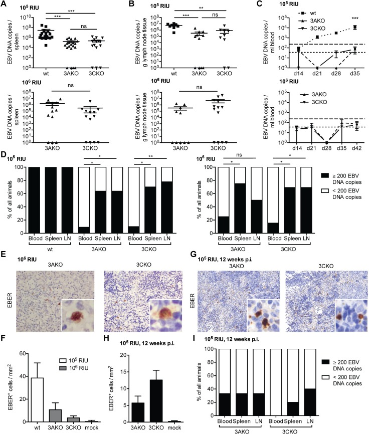
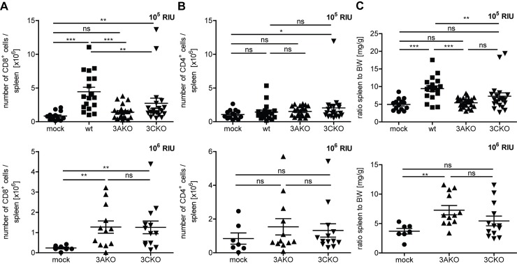
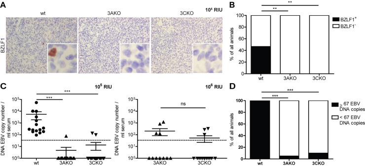
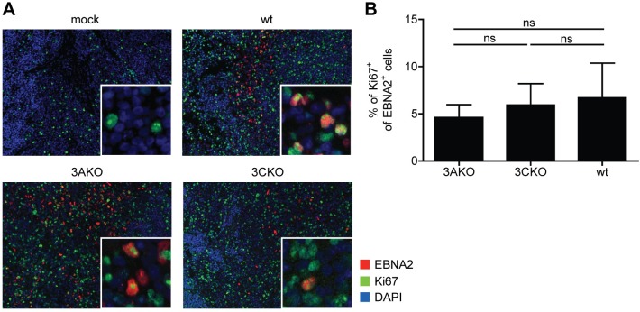
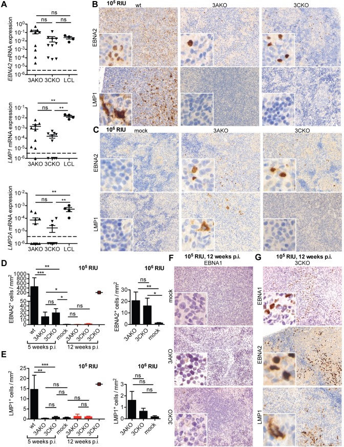
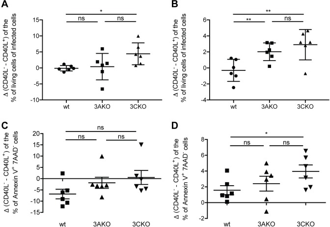
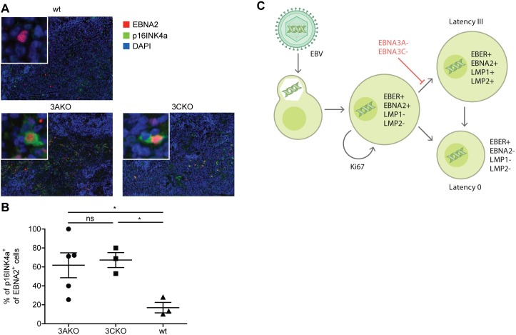
The oncogenic Epstein Barr virus (EBV) infects the majority of the human population and usually persists within its host for life without symptoms. The EBV oncoproteins nuclear antigen 3A (EBNA3A) and 3C (EBNA3C) are required for B cell transformation in vitro and are expressed in EBV associated immunoblastic lymphomas in vivo. In order to address the necessity of EBNA3A and EBNA3C for persistent EBV infection in vivo, we infected NOD-scid γcnull mice with reconstituted human immune system components (huNSG mice) with recombinant EBV mutants devoid of EBNA3A or EBNA3C expression. These EBV mutants established latent infection in secondary lymphoid organs of infected huNSG mice for at least 3 months, but did not cause tumor formation. Low level viral persistence in the absence of EBNA3A or EBNA3C seemed to be supported primarily by proliferation with the expression of early latent EBV gene products transitioning into absent viral protein expression without elevated lytic replication. In vitro, EBNA3A and EBNA3C deficient EBV infected B cells could be rescued from apoptosis through CD40 stimulation, mimicking T cell help in secondary lymphoid tissues. Thus, even in the absence of the oncogenes EBNA3A and 3C, EBV can access a latent gene expression pattern that is reminiscent of EBV persistence in healthy virus carriers without prior expression of its whole growth transforming program.


Conflict of interest statement

The authors have declared that no competing interests exist.

Figures

Fig 1
Fig 1. EBV persists without EBNA3A or EBNA3C in vivo.
(A) Splenic endpoint viral DNA load and (B) viral DNA load per gram lymph node tissue determined by qPCR of huNSG mice infected with either 105 RIU of wt, 3AKO or 3CKO for 5 weeks (spleen: n = 18-21/group, lymph node: n = 8-11/group) or 106 RIU of 3AKO or 3CKO for 6 weeks (spleen: n = 13-14/group, lymph node: n = 12-13/group). Values for mice in which no viral DNA was detected are plotted on the X-axis. (C) Blood DNA viral load over time determined by qPCR of huNSG mice infected with either 105 RIU of wt, 3AKO or 3CKO 5 weeks p.i. (n = 18-21/group) or 106 RIU of 3AKO or 3CKO 6 weeks p.i. (n = 13-14/group). Horizontal dashed line indicates the viral load of 3 times the lower limit of quantification (LLOQ). Horizontal dotted line indicates the LLOQ. (D) Frequency of huNSG mice infected with 105 RIU of 3AKO, 3CKO or wt 5 weeks p.i. or 106 RIU of 3AKO or 3CKO 6 weeks p.i. with EBV DNA copies above (≥200) or below (<200) 3 times the LLOQ in either the blood, the spleen or lymph nodes (n = 10-13/group) as determined by qPCR. Pooled data from 4 low and 2 high infectious dose experiments. *P < 0.05, **P < 0.01, Fisher’s exact test. (E) EBER-ISH (original magnification, 400x) of splenic sections from huNSG mice infected with 106 RIU of 3AKO or 3CKO 6 weeks p.i.. (G) EBER-ISH (original magnification, 200x) of splenic sections from huNSG mice infected with 105 RIU of 3AKO or 3CKO 12 weeks p.i.. (F, H) Quantification of EBER+ cells/mm2 of E (n = 10-13/group) and G (n = 3-5/group) respectively and of huNSG mice infected with 105 RIU of wt 5 weeks p.i. (n = 4/group) or mock (n = 3-8/group). (I) Frequency of huNSG mice infected with 105 RIU of 3AKO or 3CKO 12 weeks p.i., with EBV DNA copies above (≥200) or below (<200) 3 times the LLOQ in either the blood, the spleen or lymph nodes (n = 3-5/group) as determined by qPCR. Pooled data from 2 experiments. (A-C) Pooled data from 4 low and 2 high infectious dose experiments are displayed with mean ± SEM. **P < 0.01, ***P < 0.001, two-way ANOVA with Bonferroni correction for blood viral load and Mann-Whitney U test for splenic viral load, lymph node viral load and EBER-ISH.
Fig 2
Fig 2. EBNA3A or EBNA3C deficient EBV infection causes CD8+ T cell expansion.
(A) The number of splenic CD8+ T cells and (B) splenic CD4+ T cells of huNSG mice infected with either 105 RIU of wt, 3AKO or 3CKO 5 weeks p.i. (n = 14-21/group) or 106 RIU of 3AKO or 3CKO 6 weeks p.i. (n = 7-13/group) or non-infected control (mock) huNSG mice was determined by flow cytometry, applying the determined frequency to the spleen cell count. (C) The ratio of spleen to body weight (BW) of individual huNSG mice infected with either 105 RIU of wt, 3AKO or 3CKO 5 weeks p.i. (n = 14-21/group) or 106 RIU of 3AKO or 3CKO 6 weeks p.i. (n = 7-13/group) or non-infected control (mock) huNSG mice is depicted. (A-C) Pooled data from 4 low and 2 high infectious dose experiments with mean ± SEM. *P < 0.05, **P < 0.01, ***P < 0.001, Mann-Whitney U test.
Fig 3
Fig 3. EBNA3A and EBNA3C deficient EBV persist without elevated lytic replication.
(A) Representative immunohistochemistry staining for BZLF1 (original magnification, 400x) in splenic sections of huNSG mice infected with 105 RIU of wt, 3AKO or 3CKO 5 weeks p.i.. (B) Frequency of huNSG mice infected with 105 RIU of 3AKO, 3CKO or wt 5 weeks p.i. with BZLF1+ or BZLF1- cells in splenic sections as determined by immunohistochemistry. (C) DNA EBV genome copies determined by qPCR in serum of animals infected with either 105 RIU of wt, 3AKO or 3CKO 5 weeks p.i. (n = 15-20/group) or 106 RIU of 3AKO or 3CKO 6 weeks p.i. (n = 11-14/group). Values for mice in which no viral DNA was detected in the serum are plotted on the X-axis. Horizontal dotted line indicates the LLOQ. Pooled data from 4 low and 2 high infectious dose experiments represented with the mean ± SEM. ***P < 0.001, Mann-Whitney U test. (D) Frequency of huNSG mice infected with 105 RIU of 3AKO, 3CKO or wt 5 weeks p.i. with EBV DNA copies above (≥67) or below (<67) the LLOQ in the serum (n = 15-20/group) as determined by qPCR. (B, D) Pooled data from 4 experiments. ***P < 0.001, **P < 0.005, Fisher’s exact test.
Fig 4
Fig 4. EBV infected cells proliferate without EBNA3A or EBNA3C in vivo.
(A) Representative immunofluorescence staining for EBNA2 (red), Ki67 (green) and DAPI (blue) (original magnification, 200x) in splenic sections of huNSG mice infected with wt, 3AKO or 3CKO 5 or 6 weeks p.i.. (B) Quantification of A with the frequency of Ki67+ EBNA2+ cells of all EBNA2+ cells (n = 4-15/group). Pooled data from 2 experiments with mean ± SEM, Mann-Whitney U test.
Fig 5
Fig 5. EBNA3A and EBNA3C deficient EBV persist without viral protein expression.
(A) Relative EBNA2, LMP1 and LMP2A mRNA expression normalized to GAPDH in purified splenic B cells from huNSG mice infected with 106 RIU of 3AKO or 3CKO 6 weeks p.i. (n = 9-12/group) and LCL from 4 different donors. The horizontal dashed lines indicate the detection limits of the RT-qPCR assays. Values for mice in which no LMP1 transcripts were detected are plotted on the X-axis. (B, C, G) Staining for EBNA2 ((B, C) upper column, original magnification, 200x) or LMP1 ((B, C), lower column, original magnification, 200x) and (D, E) the quantification of EBNA2+ or LMP1+ cells/mm2 in splenic section of huNSG mice infected with either (B, D, E) 105 RIU of wt, 3AKO or 3CKO 5 weeks p.i. (n = 17-20/group) indicated by black bars, (D, E, G) 105 RIU of 3AKO or 3CKO 12 weeks p.i. (n = 3-5/group) indicated by red bars/square or (C-E) 106 RIU of 3AKO or 3CKO 6 weeks p.i. (n = 11-13/group) or non-infected control (mock) huNSG mice. (F, G) Staining for EBNA1 (original magnification, 200x) in splenic sections of huNSG mice infected with 3AKO or 3CKO 12 weeks p.i. or non-infected control (mock) huNSG mice. (A, D, E) Pooled data from 4 and 2 experiments represented with the mean ± SEM, **P < 0.01, ***P < 0.001, Mann-Whitney U test.
Fig 6
Fig 6. CD40 stimulation rescues EBNA3A and EBNA3C deficient EBV infected cells in vitro.
Purified human B cells were infected with wt, 3AKO or 3CKO and cultured with irradiated fibroblasts either expressing CD40L or not (n = 6). (A, B) The change in the frequency of living infected cells (GFP+) cells between the culture condition with CD40L or without CD40L for wt, 3AKO or 3CKO infected B cells (A) 2 and (B) 3 weeks p.i., determined with 7AAD life-dead stain by flow cytometry. (C, D) The change in the frequency of early apoptotic Annexin V+ 7AAD- cells between the culture condition with CD40L or without CD40L for wt, 3AKO or 3CKO infected B cells (C) 2 and (D) 3 weeks p.i., determined by flow cytometry. (A-D) Pooled data from 3 experiments represented with the mean ± SEM, *P < 0.05, **P < 0.01, ***P < 0.001, paired t-test.
Fig 7
Fig 7. Elevated p16INK4a expression in EBV infected cells without EBNA3A or 3C in vivo.
(A) Representative immunofluorescence staining for EBNA2 (red), p16INK4a (green) and DAPI (blue) (original magnification, 200x) in splenic sections of huNSG mice infected with wt, 3AKO or 3CKO 5 or 6 weeks p.i.. (B) Quantification of A with the frequency of p16INK4a+ EBNA2+ cells of all EBNA2+ cells (n = 3-5/group). Pooled data from 2 experiments with mean ± SEM, unpaired t-test with Welch's correction. (C) Schematic representation of B cell infection by EBV in humanized mice.

References

    1. Epstein MA, Achong BG, Barr YM. Virus particles in cultured lymphoblasts from Burkitt's lymphoma. Lancet. 1964;1:702–3. - PubMed
    1. Epstein MA, Henle G, Achong BG, Barr YM. Morphological and biological studies on a virus in cultured lymphoblasts from Burkitt's lymphoma. J Exp Med. 1964;121:761–70. - PMC - PubMed
    1. Nilsson K, Klein G, Henle W, Henle G. The establishment of lymphoblastoid lines from adult and fetal human lymphoid tissue and its dependence on EBV. Int J Cancer. 1971;8(3):443–50. - PubMed
    1. Cesarman E. Gammaherpesviruses and lymphoproliferative disorders. Annu Rev Pathol. 2014;9:349–72. doi: 10.1146/annurev-pathol-012513-104656 - DOI - PubMed
    1. Babcock JG, Hochberg D, Thorley-Lawson AD. The expression pattern of Epstein-Barr virus latent genes in vivo is dependent upon the differentiation stage of the infected B cell. Immunity. 2000;13(4):497–506. - PubMed

Publication types

Substances