Review
. 2018 Jul;28(4):e1973.
doi: 10.1002/rmv.1973. Epub 2018 Apr 30.

Autophagy: The multi-purpose bridge in viral infections and host cells

Affiliations
Review

Autophagy: The multi-purpose bridge in viral infections and host cells

Asghar Abdoli et al. Rev Med Virol. 2018 Jul.

Abstract

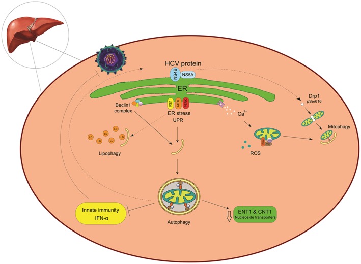
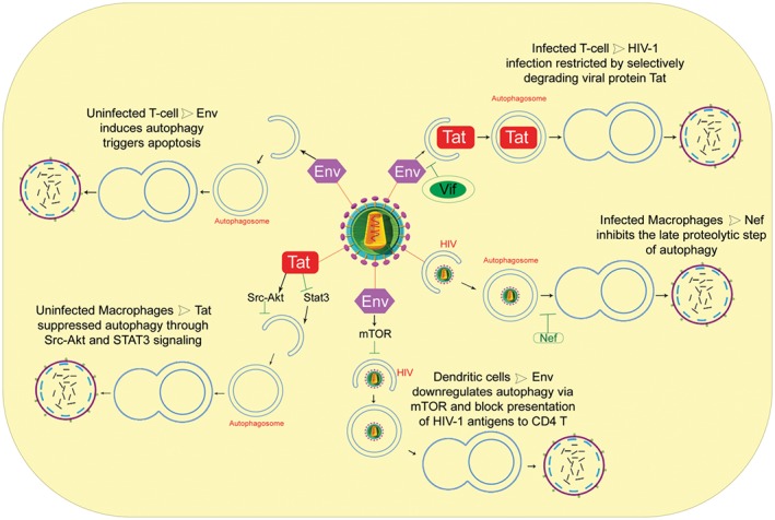
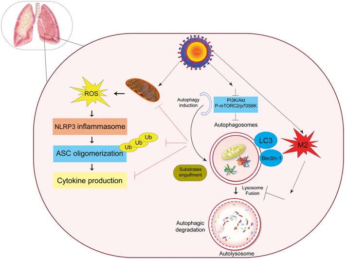
Autophagy signaling pathway is involved in cellular homeostasis, developmental processes, cellular stress responses, and immune pathways. The aim of this review is to summarize the relationship between autophagy and viruses. It is not possible to be fully comprehensive, or to provide a complete "overview of all viruses". In this review, we will focus on the interaction of autophagy and viruses and survey how human viruses exploit multiple steps in the autophagy pathway to help viral propagation and escape immune response. We discuss the role that macroautophagy plays in cells infected with hepatitis C virus, hepatitis B virus, rotavirus gastroenteritis, immune cells infected with human immunodeficiency virus, and viral respiratory tract infections both influenza virus and coronavirus.

Keywords: autophagy; host cell; interaction; virus.


Conflict of interest statement

None declared.

Figures

Figure 1
Figure 1
The cross‐talk between autophagy and HCV. The hepatitis C virus (HCV) induces autophagy to enhance virus replication rather than to block it. After HCV decapsidation, the virus genome is transferred to endoplasmic reticulum (ER) and initiates replication in the ER‐derived membranous web as the HCV replication platform. HCV‐induced ER stress disrupts ER function and results in accumulation of unfolded proteins in the ER. Cells adapt to ER stress by triggering an unfolded protein response (UPR) to restore ER homeostasis. The UPR signaling pathway includes 3 central elements, PKR‐like ER kinase (PERK), ATF6, and IRE1. The UPR could stimulate an autophagy response. Under these conditions, calcium released from the ER stimulates impaired mitochondrial activity connect to excessive production of ROS. Dysfunctional mitochondria are engulfed for degradation via mitophagy. In addition, HCV promoted dynamin‐related protein 1‐mediated mitochondrial fission protein 1 (Drp1) leads to mitophagy. HCV replication takes place in close proximity to lipid droplets (LDs), the neutral lipid storage organelles, and this closeness enables the downstream viral morphogenetic events. A selective autophagy for lipids (lipophagy) protects cells from an excessive lipid accumulation elicited by HCV. Upon HCV infection, the host stimulates the antiviral interferon‐mediated frontline. HCV employs autophagy to escape host innate immune surveillance by inhibiting IFNα production. Furthermore, HCV replication reduced ribavirin uptake, due to reduced expression of the nucleoside transporters ENT1 and CNT1
Figure 2
Figure 2
Schematic overview of the autophagy and HIV‐1 in target cells. Autophagy, as a cell‐autonomous innate defense, plays an important role against HIV‐1 infection, it restricts hiv‐1 infection by Tat degradation and limits virion production. However, HIV‐1 proteins are able to modulate autophagy pathway by different strategy at the initiation and maturation step (Tat in uninfected macrophages; Env in dendritic cells; Vif in infected T cells; Nef in infected macrophages). In particular, in infected macrophages, Nef acts as an “antiautophagic maturation factor” and blocks the autophagosome maturation step. In uninfected CD4 T cells, Env‐mediated autophagy leads to apoptotic cell death
Figure 3
Figure 3
Regulatory signaling of autophagy during influenza A virus infection. Influenza A virus as inducer of the NLRP3 inflammasome causes mitochondrial damage, which leads to the release of reactive oxygen species (ROS). NLRP3 forms an inflammasome complex with ASC and induces the production of inflammatory cytokines. Influenza A virus also binds to Beclin1 by viral M2 protein. It upregulates the expression of several autophagy related genes, which can increase the autophagic flux, and M2 also contains an LC3‐interacting region (LIR), which is required for influenza virus subversion of autophagy which leads to LC3 redistribution to the plasma membrane in infected cells

Similar articles

Cited by

References

    1. Cao Y, Klionsky DJ. Physiological functions of Atg6/Beclin 1: a unique autophagy‐related protein. Cell Res. 2007;17(10):839‐849. - PubMed
    1. Yang Z, Klionsky DJ. Eaten alive: a history of macroautophagy. Nat Cell Biol. 2010;12(9):814‐822. - PMC - PubMed
    1. Levine B, Klionsky DJ. Development by self‐digestion: molecular mechanisms and biological functions of autophagy. Dev Cell. 2004;6(4):463‐477. - PubMed
    1. Eskelinen E‐L. Maturation of autophagic vacuoles in mammalian cells. Autophagy. 2005;1(1):1‐10. - PubMed
    1. Axe EL, Walker SA, Manifava M, et al. Autophagosome formation from membrane compartments enriched in phosphatidylinositol 3‐phosphate and dynamically connected to the endoplasmic reticulum. J Cell Biol. 2008;182(4):685‐701. - PMC - PubMed

LinkOut - more resources