Review
. 2018 Jul 8:41:475-499.
doi: 10.1146/annurev-neuro-080317-061756. Epub 2018 Apr 30.

The Dynamic Basis of Respiratory Rhythm Generation: One Breath at a Time

Affiliations
Review

The Dynamic Basis of Respiratory Rhythm Generation: One Breath at a Time

Jan-Marino Ramirez et al. Annu Rev Neurosci. .

Abstract

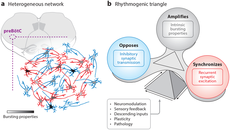
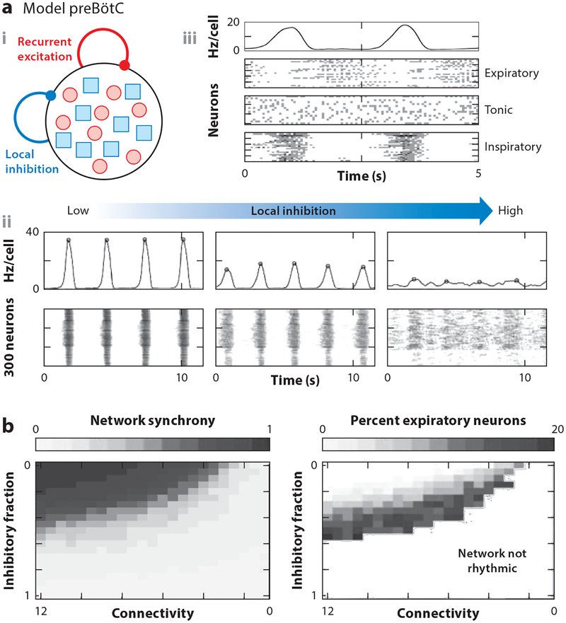
Rhythmicity is a universal timing mechanism in the brain, and the rhythmogenic mechanisms are generally dynamic. This is illustrated for the neuronal control of breathing, a behavior that occurs as a one-, two-, or three-phase rhythm. Each breath is assembled stochastically, and increasing evidence suggests that each phase can be generated independently by a dedicated excitatory microcircuit. Within each microcircuit, rhythmicity emerges through three entangled mechanisms: ( a) glutamatergic transmission, which is amplified by ( b) intrinsic bursting and opposed by ( c) concurrent inhibition. This rhythmogenic triangle is dynamically tuned by neuromodulators and other network interactions. The ability of coupled oscillators to reconfigure and recombine may allow breathing to remain robust yet plastic enough to conform to nonventilatory behaviors such as vocalization, swallowing, and coughing. Lessons learned from the respiratory network may translate to other highly dynamic and integrated rhythmic systems, if approached one breath at a time.

Keywords: breathing; coupled oscillators; excitation/inhibition balance; microcircuits; rhythm generation; synchronization.


Figures

Figure 1
Figure 1
Respiratory phases can act independently, reconfigure, and exhibit stochasticity. (a) Respiratory-related electromyographic activity of the latissimus dorsi (LD) muscle during a single breath in a trained classical singer performing an exercise from “Una voce poco fa” from The Barber of Seville by Gioachino Rossini. Note the repeated bouts of LD activity during postinspiration (arrows in expanded view) that correlate with vibrato visible in the corresponding sonogram (figure adapted with permission from Watson et al. 2012). (b) Experiment from an in situ arterially perfused rat preparation showing recordings of activity from the phrenic (Ph) nerve, innervating the diaphragm; cervical vagal nerve (cVN), containing a branch that innervates laryngeal muscles; and hypoglossal (XII) nerve, innervating the tongue, under normoxic and hypoxic conditions. During normoxia, postinspiratory and preinspiratory activity are prominent in cVN and XII recordings, respectively (arrows). But activity in the cVN and XII becomes primarily inspiratory during hypoxia, demonstrating the ability of the respiratory network to reconfigure from a three-phase rhythm to a one-phase rhythm (figure adapted with permission from Paton et al. 2006). (c) Plethysmography recording from an awake behaving mouse reveals stochasticity in breath waveforms.
Figure 2
Figure 2
The ventral respiratory column (VRC) in the medulla provides the basis for respiratory rhythm generation. The VRC contains (from rostral to caudal) the retrotrapezoid nucleus/parafacial respiratory group (RTN/pFRG), encompassing the ventral borders of the facial nucleus (VII); the postinspiratory complex (PiCo), caudal to VII and dorsomedial to the nucleus ambiguus (NA); the Bötzinger complex (BötC) and preBötzinger complex (preBötC), ventromedial to the NA; and the rostral and caudal ventral respiratory groups (rVRG and cVRG, respectively), dorsal to the lateral reticular nucleus (LRN). The borders of VRC compartments are indistinct. However, genetic and molecular markers that characterize the rhythmic regions of the VRC have been described. (a) In the preBötC, rhythmic excitatory neurons are derived from precursors that express the transcription factor Dbx1 during development (white arrowheads mark Dbx1 neurons colabeled with the neuron-specific AAV-hSYN-GFP). Neurons along the inter-reticular zone (IRt), including XII premotor neurons, and some glia (gray arrowheads), are also derived from Dbx1-expressing cells (figure adapted from Kottick et al. 2017 with permission). (b) PiCo neurons are defined by coexpression of vesicular glutamate transporter 2 (Vglut2) and choline acetyltransferase (ChAT). White arrows specify neurons containing nuclei expressing Cre (under control of the Vglut2 promoter) and ChAT within the surrounding cytoplasm (figure adapted from Anderson et al. 2016). (c) The RTN/pFRG contains neurons that express Phox2b but not tyrosine hydroxylase (TH) or ChAT (figure adapted from Stornetta et al. 2006 with permission). Other abbreviations: AAV-hSYNGFP, adeno-associated virus-human synapsin 1-green fluorescent protein; DAPI, 4′,6-diamidino-2-phenylindole; ECu, external cuneate nucleus; icp, inferior cerebellar peduncle; IO, inferior olive; ml, medial lemniscus; MVe, medial vestibular nucleus; py, pyramidal tract; Sp5, spinal trigeminal tract; SpVe, spinal vestibular nucleus; XII, hypoglossal motor nucleus.
Figure 3
Figure 3
Interactions in a heterogeneous network give rise to a rhythmogenic triangle. (a) The isolated preBöTc network contains excitatory and inhibitory neurons with varied degrees of burstiness. (b) Network synchrony is regulated by the rhythmogenic triangle: Glutamatergic synaptic transmission is essential for synchronization, intrinsic membrane bursting conductances enhance spiking and amplify synchronization, and concurrent inhibition opposes synchronization and increases variability. The balance of these components can be tuned to adapt breathing for specific behaviors and states by, for example, neuromodulation, sensory feedback, descending inputs, and plasticity; whereas pathology can disrupt this balance, leading to breathing disturbances that can be behavior- and state-dependent. Abbreviation: preBötC, preBötzinger complex.
Figure 4
Figure 4
Respiratory rhythm-generating networks contain neurons with variable firing patterns and stochastic activity. (a) Multielectrode array recordings from the preBöTc (77 experiments with 918 units) reveal that most neurons are active in phase with inspiration. (b) Spike rasters of 11 simultaneously recorded inspiratory neurons over 150 respiratory cycles demonstrating substantial variability in the amount of spiking activity, phase modulation, burst onset, and burst shape and duration between neurons. Red arrow indicates preBötC population burst onset. Integrated spiking activity of each neuron is shown on the right. Panels a and b adapted from Carroll et al. 2013. (c) Intracellular recording of a single inspiratory neuron over 10 respiratory cycles illustrating the stochasticity of burst onset relative to preBötC population activity (red arrow). Abbreviation: preBötC, preBötzinger complex.
Figure 5
Figure 5
Network connectivity and bursting properties influence stochasticity and the transition to a population burst. (a) Comparison between fully and sparsely connected preBötC models containing excitatory neurons and bursting currents. Sparse network connectivity reproduces the burst onset variability (arrows) characteristic of the preBöTc (see Figure 4b,c). Sparsity can also prohibit the network from fully synchronizing in some cycles, resulting in a weak population burst (arrow in integrated model activity). (b) Within the network, bursting currents amplify excitatory synaptic drive and facilitate transmission of preBötC population activity to motor output. The effect of bursting currents can be observed by comparing burst activity of a neuron under control conditions and in the presence of a slight hyperpolarizing current in which weak inspiratory activity is no longer able to translate synaptic drive into a burst (illustrated in the expanded overlay) and preBöTc activity is not successfully transmitted to XII motor output (left). The amplifying influence of bursting properties is also indicated by the ability to prematurely terminate an ongoing burst with a brief hyperpolarizing current pulse (right). Figures adapted with permission from Ramirez & Richter (1996). (c) The presence of persistent sodium current (INaP) and calcium-activated nonselective cation current (ICAN) gives rise to a gradient of burstiness among preBötC neurons. Some neurons, called intrinsic or endogenous bursting neurons, continue to burst in the absence of synaptic interactions. Abbreviations: preBötC, preBötzinger complex; XII, hypoglossal motor nucleus.
Figure 6
Figure 6
Neurons receive concurrent excitation and inhibition from within the isolated preBötC, a critical feature regulating burst shape, phase, and excitability. (a) Voltage clamp recording from an inspiratory preBöTc neuron showing concurrent EPSCs (red) and IPSCs (blue) synchronized with integrated preBötC population activity (gray). (b) Activity of optogenetically identified excitatory Dbx1- and inhibitory Vgat-expressing neurons during preBötC population bursts. (c) Loss of synaptic inhibition transforms the augmenting burst shape of inspiratory preBötC neurons into a decrementing shape; expiratory neurons become inspiratory (red and blue arrows reflect the relative influence of excitatory and inhibitory synaptic inputs, respectively). With glutamatergic synaptic transmission blocked (CNQX), tonic activity in some inspiratory neurons is transformed into endogenous bursting (figure adapted from Tryba et al. 2003). Abbreviations: E, excitation; EPSCs, excitatory postsynaptic currents; I, inhibition; IPSCs, inhibitory postsynaptic currents; preBötC, preBotzinger complex.
Figure 7
Figure 7
The preBötC network reconfigures in hypoxia. (a) Population activity in the isolated preBötC at baseline and during the transition to hypoxia. Fictive eupneic bursts transition to fictive gasping. Averaged eupnea (black) and gasping (red) burst waveforms illustrating changes in burst rise time and duration are shown on the right. (b) Multi-array recordings reveal that, although the average activity of the preBötC is reduced in hypoxia, individual respiratory elements have varied responses to hypoxia. Some exhibit increased mean firing rate, whereas others are completely suppressed (black dots). (c) Graphic representation of preBötC network configurations in normoxia and hypoxia. Respiratory elements are represented as circles with a diameter proportional to the number of functional connections as determined by cross-correlation analysis. Significant correlations are represented as connecting lines, with the weight of the line reflecting the strength of each connection. Note that reconfiguration of the network involves changes in the number and strength of the connections between individual elements, but that the overall number of elements and connections within the network remains relatively unchanged (panels b and c adapted with permission from Nieto-Posadas et al. 2014). (d) Quantification of EPSCs and IPSCs from inspiratory preBötC neurons during the transition to hypoxia demonstrates that changes in activity are not evenly distributed among excitatory and inhibitory neurons. (e) Activity of an expiratory preBötC neuron during network reconfiguration in hypoxia. Note the loss of inhibition during inspiration (blue arrows), resulting in a phase transition from expiratory activity during fictive eupnea to inspiratory activity during fictive gasping. Asterisk indicates fictive sigh bursts. Abbreviations: EPSCs, excitatory postsynaptic currents; IPSCs, inhibitory postsynaptic currents; preBötC, preBötzinger complex.
Figure 8
Figure 8
The preBötC operates on the edge of synchrony. (a) Representation of increasing amounts of heterogeneity included in preBöTc network models. (a, i) Schematic of a model preBötC examining the role of inhibitory neurons in rhythm generation. (a, ii) Integrated rhythmic model preBötC activity and spike rasters with an increasing fraction of inhibitory neurons. (a, iii) Represents the same network parameters shown in the middle panels of a,ii, demonstrating the emergence of neurons with expiratory activity. (b) As the fraction of inhibitory neurons and the sparsity of network connectivity increase, network synchrony is reduced and the percentage of neurons with expiratory activity increases to 20%, at which point synchrony is lost and the network is no longer rhythmic (figures adapted with permission from Harris et al. 2017). Note that approximately 25–50% of the neurons in the preBötC are inhibitory (Morgado-Valle et al. 2010, Winter et al. 2009) and approximately 10% of neurons are expiratory (see Figure 3) (Carroll et al. 2013), suggesting the preBötC operates near the edge of synchrony. Abbreviation: preBötC, preBötzinger complex.
Figure 9
Figure 9
Coupled rhythmogenic networks are required to generate a multiphase breathing rhythm.(a, i) Computational model of two rhythmic networks, containing recurrent excitation and local inhibition, coupled by long-range inhibition. (a, ii) Comparison of simulated rhythmic activity in networks with strong local inhibition and weak long-range inhibition (top), and in networks with weak local inhibition and strong long-range inhibition (bottom) (figure adapted with permission from Harris et al. 2017). (b) Schematic representation of the triple oscillator hypothesis, in which the preBötC, PiCo, and, under high metabolic demand, RTN/pFRG interact via mutually inhibitory and excitatory connections (connections indicated by dashed lines are speculative). Inhibition dominates under normal conditions, resulting in a multiphase rhythm; but like the rhythmogenic triangle, the balance of excitation and inhibition between oscillators can be shifted to facilitate phase reconfiguration such as during hypoxia (see Figure 1b). Abbreviations: PiCo, postinspiratory complex; preBötC, preBötzinger complex; RTN/pFRG, retrotrapezoid nucleus/parafacial respiratory group.

References

    1. Abdala AP, Toward MA, Dutschmann M, Bissonnette JM, Paton JF. 2016. Deficiency of GABAergic synaptic inhibition in the Kölliker-Fuse area underlies respiratory dysrhythmia in a mouse model of Rett syndrome. J. Physiol 594:223–37 - PMC - PubMed
    1. Alheid GF, McCrimmon DR. 2008. The chemical neuroanatomy of breathing. Respir. Physiol. Neurobiol 164:3–11 - PMC - PubMed
    1. Anderson TM, Garcia AJ 3rd, Baertsch NA, Pollak J, Bloom JC, et al. 2016. A novel excitatory network for the control of breathing. Nature 536:76–80 - PMC - PubMed
    1. Anderson TM, Ramirez JM. 2017. Respiratory rhythm generation: triple oscillator hypothesis. F1000Research 6:139. - PMC - PubMed
    1. Bacak BJ, Kim T, Smith JC, Rubin JE, Rybak IA. 2016. Mixed-mode oscillations and population bursting in the pre-Bötzinger complex. eLife 5:e13403. - PMC - PubMed

Publication types

LinkOut - more resources