Review
. 2018 Jul;43(7):517-532.
doi: 10.1016/j.tibs.2018.04.002. Epub 2018 Apr 27.

Amazing Diversity in Biochemical Roles of Fe(II)/2-Oxoglutarate Oxygenases

Affiliations
Review

Amazing Diversity in Biochemical Roles of Fe(II)/2-Oxoglutarate Oxygenases

Caitlyn Q Herr et al. Trends Biochem Sci. 2018 Jul.

Abstract

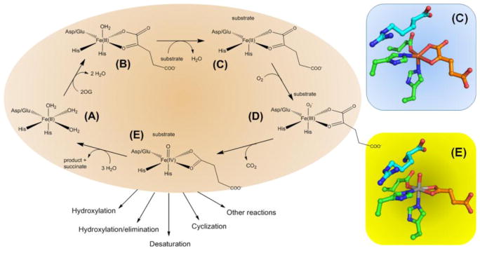
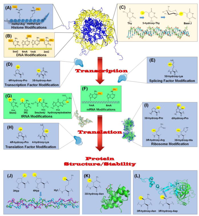
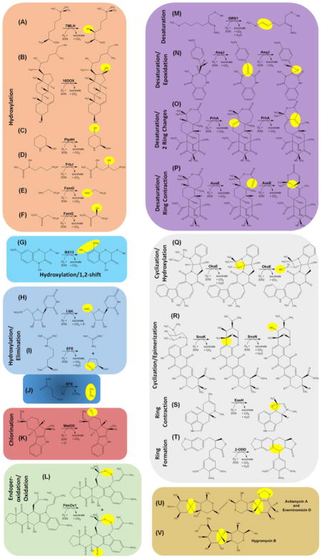
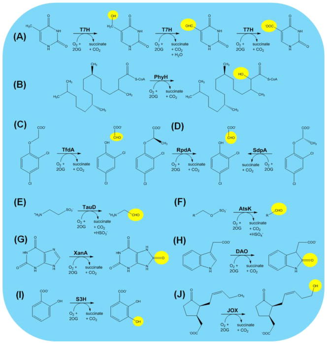
Since their discovery in the 1960s, the family of Fe(II)/2-oxoglutarate-dependent oxygenases has undergone a tremendous expansion to include enzymes catalyzing a vast diversity of biologically important reactions. Recent examples highlight roles in controlling chromatin modification, transcription, mRNA demethylation, and mRNA splicing. Others generate modifications in tRNA, translation factors, ribosomes, and other proteins. Thus, oxygenases affect all components of molecular biology's central dogma, in which information flows from DNA to RNA to proteins. These enzymes also function in biosynthesis and catabolism of cellular metabolites, including antibiotics and signaling molecules. Due to their critical importance, ongoing efforts have targeted family members for the development of specific therapeutics. This review provides a general overview of recently characterized oxygenase reactions and their key biological roles.

Keywords: Nonheme iron oxygenase; biodegradation; biosynthesis; chromatin modification; transcription; translation.


Figures

Figure 1
Figure 1
Generalized Mechanism of 2OG-Dependent Oxygenases. (A) Active site metallocenter with Fe(II) coordinated by a 2-His-1-carboxylase motif. (B) 2OG-bound protein. (C) Creation of an O2-binding site in the substrate- and 2OG-bound state. (D) Fe(III)-superoxo species. (E) Ferryl intermediate that is common to all further chemistry. Also depicted are two views of the VioC active site: (C) Fe(II)-containing protein with bound L-Arg and 2OG (PDB access code 6ALM) and (E) vanadyl-containing protein (as a mimic of the ferryl species) with bound L-Arg and succinate (PDB access code 6ALR).
Figure 2
Figure 2
Relationship of 2OG-Dependent Oxygenases to the Central Dogma. Chromatin regulation involves methylation of (A) specific Lys and Arg residues in the N-terminal tails of histones and (B) several types of bases in DNA. Demethylases and other 2OG-dependent oxygenases function to reverse these modifications. (C) Base J is a DNA modification found in certain kinetoplasts. Selected thymidine bases (Thy) are modified by a 2OG-dependent hydroxylase to form 5-hydroxy-Thy, then glycosylated by a separate enzyme. (D) Regulated RNA synthesis involves transcription factors, such as HIF, modified by 2OG-dependent hydroxylases. (E) An oxygenase also modifies a splicing factor as part of the maturation of precursor RNA. (F) 2OG-dependent enzymes remove methylation marks in mRNA. (G) Representatives of these enzymes demethylate, hydroxylate, and hypermodify bases in tRNA. (H) Translation factors undergo hydroxylations catalyzed by 2OG-dependent oxygenase. (I) Several hydroxylations are introduced into ribosomal proteins by these enzymes. In addition, other proteins are hydroxylated related to structure/stability. (J) Collagen contains 4R-hydroxy-Pro (4Hyp), 3S-hydroxy-Pro (3Hyp), and 5R-hydroxy-Lys (Hyl) residues synthesized by 2OG-dependent hydroxylases. The structure is shown for a collagen model peptide (PDB access code 3ABN) that is rich in Pro and Gly residues and contains 4Hyp (arrow); three peptides (depicted in green, cyan, and magenta) form a triple-helical structure. (K) Some ankyrin repeat domain (ARD) proteins undergo hydroxylation at the 3S position of Asn residues, leading to protein stabilization. The structure shown (PDB access code 2ZGD) is from a synthetic consensus sequence hydroxylated by FIH. (L) Activated factor IX (PDB access code 1PFX) has a large catalytic chain (green) and a small chain (cyan) containing an EGF domain with a 3R-hydroxy-Asp residue (arrow). Also shown is the structure of 3R-hydroxy-Asn. DNA bases are shown in yellow, RNA components are in green, and protein sidechains are in blue. The methylation sites targeted for hydroxylation (A, B, and F) and the introduced hydroxylations (C, D, E, G, H, I, J, K, and L) are highlighted in brown and yellow, respectively.
Figure 3
Figure 3
Representative Biosynthetic Reactions of 2OG-Dependent Oxygenases. (A–F) Selected hydroxylation reactions. (G) A hydroxylation reaction coupled with a 1,2-shift of a methoxy group. (H–I) Hydroxylation reactions followed by spontaneous elimination reactions. The enzyme catalyzing reaction (I) also catalyzes reaction (J) (see Box 2). (K) A chlorination reaction. (L) An endoperoxide-forming reaction, along with an oxidation reaction. (M–P) Desaturation reactions, in most cases accompanied by ring formation or ring rearrangement reactions. (Q–T) Additional ring transformation reactions. (U–V) Examples of orthoether linkages and methylenedioxy groups synthesized by 2OG-dependent oxygenases. See text for details.
Figure 4
Figure 4
Representative Biodegradative Reactions of 2OG-Dependent Oxygenases. (A) Thymine 7-hydroxylase (T7H). (B) Phytanoyl-CoA hydroxylase (PhyH). (C) 2,4-Dichlorophenoxyacetic acid hydroxylase (TfdA). (D) (R)- and (S)-specific dichlorophenoxypropionic acid hydroxylases (RdpA and SdpA). (E) Taurine hydroxylase (TauD). (F) Alkylsulfate hydroxylase (AtsK). (G) Xanthine hydroxylase (XanA). (H) Auxin dioxygenase (DAO). (I) Salicyclic acid 3-hydroxylase (S3H). (J) Jasmonic acid oxygenase (JOX).

References

    1. Hausinger RP. Biochemical diversity of 2-oxoglutarate-dependent oxygenases. In: Schofield CJ, Hausinger RP, editors. 2-Oxoglutarate-Dependent Oxygenases. Royal Society of Chemistry; 2015. pp. 1–58.
    1. Wu LF, et al. Ferrous iron and α-ketoglutarate dependent dioxygenases in the biosynthesis of microbial natural products. Biochim Biophys Acta. 2016;1864:453–470. - PubMed
    1. Islam MS, et al. 2-Oxoglutarate-dependent oxygenases. Annu Rev Biochem. 2018 in press. - PubMed
    1. Hagel JM, Facchini PJ. Expanding the roles for 2-oxoglutarate-dependent-oxygenases in plant metabolism. Natural Prod Rep. 2018 in press. - PubMed
    1. Aik WS, et al. Introduction to structural studies on 2-oxoglutarate-dependent oxygenases and related enzymes. In: Schofield CJ, Hausinger RP, editors. 2-Oxoglutarate-Dependent Oxygenases. Royal Society of Chemistry; 2015.

Publication types

MeSH terms