Review
. 2018 Sep;88(3):413-426.
doi: 10.1016/j.gie.2018.04.2352. Epub 2018 Apr 27.

Screening for esophageal squamous cell carcinoma: recent advances

Affiliations
Review

Screening for esophageal squamous cell carcinoma: recent advances

Don C Codipilly et al. Gastrointest Endosc. 2018 Sep.

Abstract

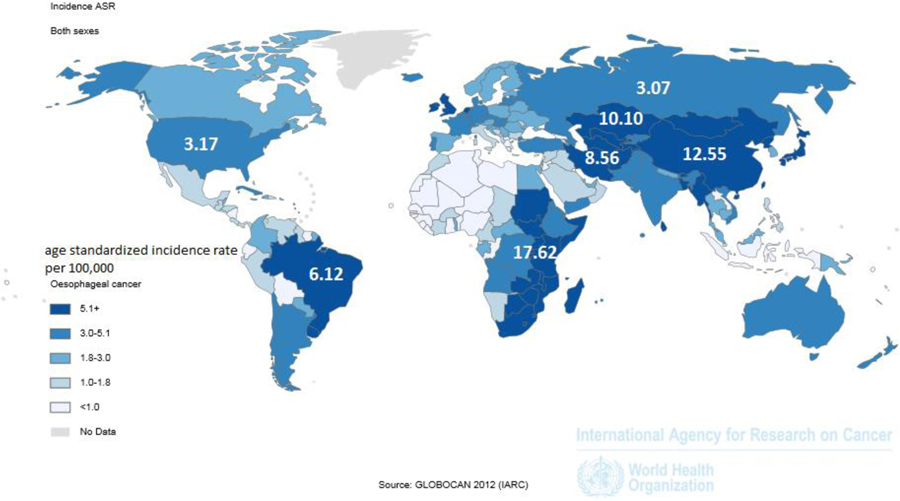
Esophageal squamous cell carcinoma (ESCC) is the most common type of esophageal cancer worldwide, with a high mortality due to advanced stage at diagnosis. Although most common in an area known as the Asian Esophageal Cancer Belt, which extends from the Caspian Sea to northern China, and in parts of Africa, high-risk populations also exist elsewhere in the world. Screening for ESCC has been practiced in a few geographic areas and high-risk populations, with varying levels of success. Esophageal squamous dysplasia is recognized as the precursor lesion for ESCC. Endoscopic screening for ESCC/esophageal squamous dysplasia is expensive and not sufficiently available in many high-risk regions. Recent advances in non-endoscopic screening enhanced by biomarker-based disease detection have raised the prospect of improved accuracy and availability of screening for esophageal squamous dysplasia and early stage ESCC. Development of a cost-effective, accurate, and well-tolerated screening test, if applied in endemic areas and high-risk populations, has the potential to reduce mortality from this deadly disease worldwide. In this review, we summarize recent developments in endoscopic and non-endoscopic screening modalities.


Conflict of interest statement

Conflicts of Interest:

Prasad G. Iyer: Research funding from Exact Sciences, C2 Therapeutics, Medtronic, Nine Point Medical

John B. Kisiel and David A. Ahlquist: Listed as co-inventors in an intellectual property development agreement with Exact Sciences and could receive future royalties.

Figures

Figure 1:
Figure 1:
The Asian and African esophageal cancer belts (in dark blue), where over 90% of cases are esophageal squamous cell carcinoma. (Reprinted with permission from Ferlay J SI, et al. GLOBOCAN 2012 v1.0, [Internet]. Lyon, France: International Agency for Research on Cancer, 2013.)
Figure 2:
Figure 2:
Original intrapapillary capillary loop (IPCL) pattern classification. The IPCL pattern classification includes two sets of diagnostic criteria. IPCL pattern classification from IPCL type I to type V-1 is used for the tissue characterization of flat lesions (red outline). IPCL pattern classification from IPCL type V-1 to type VN reflects cancer infiltration depth (blue outline). IPCL type III corresponds to borderline lesions which potentially include esophagitis or low-grade intraepithelial neoplasia. IPCL type III should be considered for endoscopic follow up. In IPCL type IV, high-grade intraepithelial neoplasia appears, and then further treatment with endoscopic mucosal resection (EMR) / endoscopic submucosal dissection is recommended. EMR/esophageal squamous dysplasia for IPCL types V-1 and V-2 should be also considered as they are definite M1 or M2 lesion with no risk of lymph node metastasis. IPCL type V3 corresponds to an M3 lesion and diagnostic EMR/esophageal squamous dysplasia should be applied as a “complete biopsy” to decide on a final treatment strategy. IPCL type VN corresponds to a “new tumor vessel” often associated with sm2 invasion with significantly increased risk of lymph node metastasis. Surgical treatment should be recommended. (Reprinted with permission from Inoue H, et al. Ann Gastroenterol. 2015 Jan-Mar; 28(1): 41–48.)
Figure 3:
Figure 3:
Endoscopic images of the same lesion of esophageal squamous dysplasia, with WLE, NBI, Magnifying NBI, and Lugol’s chromoendoscopy. a. | Endoscopic finding with conventional white light imaging. A flat reddish lesion was seen in the upper thoracic esophagus (between the arrows). At the edge of the lesion, the vascular pattern in the surrounding mucosa was interrupted. b. | Endoscopic finding with narrow band imaging of the lesion from panel a. The lesion was contrasted as darker area (arrow) and the boundary of the lesion is clearer than in white light images. c. | Endoscopic finding with magnifying narrow-band imaging. Dilated irregular microvascular pattern was clearly visualized in the lesion. A distinct demarcation line between the background mucosa and the lesion is visible (arrow). Note the color of the epithelium in the lesion is browner than the surrounding mucosa. Accordingly, endoscopic diagnosis of intramucosal squamous cell carcinoma or high-grade dysplasia can be made with high confidence. d. | Endoscopic finding using Lugol’s chromoendoscopy for the lesion depicted in panels a and b. The lesion is clearly visualized as a lighter (yellow) unstained area within the brown stained area. The boundary of the lesion can be clearly delineated with chromoendoscopy. (Reprinted with permission from Veitch AM, et al. Nat Rev Gastroenterol & Hepatol. 2015)
Figure 4:
Figure 4:
Microendoscopic imaging of squamous dysplasia. a. | Endocytoscope probe which can be passed through the instrument channel of an endoscope. b. | The border between esophageal squamous cancer (upper) and normal squamous epithelium (lower). The density and nucleus:cytoplasm ratio is much higher in cancer compared to normal epithelium. (Reprinted with permission from Kumagai Y, et al. Endoscopy 2004;36:590–594.)
Figure 5:
Figure 5:
High-resolution microendoscope Lugol’s unstained lesions (left) are imaged with HRME (“optical” biopsy) and compared with corresponding tissue biopsy (histopathologic biopsy) (original magnification, 100x). Only 1 of the 2 Lugol’s abnormal areas was neoplastic (upper panel) as determined by the imaging software, based on mean nuclear area, nuclear-to cytoplasmic ratio, nearest inter-nuclear distance, nuclear eccentricity, nuclear solidity, and the major axis of the ellipse best approximating each nucleus. (Reprinted with permission from Protano et al. Gastroenterology 2015; 149: 321–329.)
Figure 6:
Figure 6:
Balloons and sponges used in prior studies for screening of esophageal squamous cell carcinoma and/or dysplasia. (Reprinted with permission from Pan QJ, et al. Acta Cytol 2008;52:14–23.)

Comment in

References

    1. Ferlay JSI, Ervik M, Dikshit R, Eser S, Mathers C, Rebelo M, Parkin DM, Forman D, Bray F. GLOBOCAN 2012 v1.0, Cancer Incidence and Mortality Worldwide: IARC CancerBase No. 11 [Internet]. Lyon, France: International Agency for Research on Cancer, 2013.
    1. Jemal A, Bray F, Center MM, et al. Global cancer statistics. CA Cancer J Clin 2011;61:69–90. - PubMed
    1. Siegel RL, Miller KD, Jemal A. Cancer statistics, 2016. CA: A Cancer Journal for Clinicians 2016;66:7–30. - PubMed
    1. Arnold M, Soerjomataram I, Ferlay J, et al. Global incidence of oesophageal cancer by histological subtype in 2012. Gut 2015;64:381–7. - PubMed
    1. Torre LA, Siegel RL, Ward EM, et al. Global Cancer Incidence and Mortality Rates and Trends--An Update. Cancer Epidemiol Biomarkers Prev 2016;25:16–27. - PubMed

MeSH terms