Review
. 2018 Apr;16(2):85-98.
doi: 10.1016/j.gpb.2018.03.001. Epub 2018 Apr 27.

Structural Insights into N6-methyladenosine (m6A) Modification in the Transcriptome

Affiliations
Review

Structural Insights into N6-methyladenosine (m6A) Modification in the Transcriptome

Jinbo Huang et al. Genomics Proteomics Bioinformatics. 2018 Apr.

Abstract

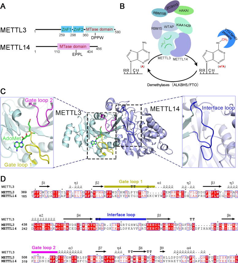
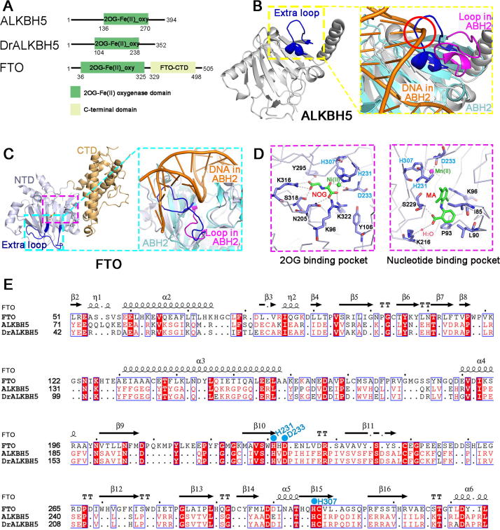
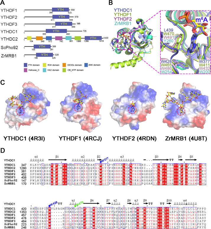
More than 100 types of chemical modifications in RNA have been well documented. Recently, several modifications, such as N6-methyladenosine (m6A), have been detected in mRNA, opening the window into the realm of epitranscriptomics. The m6A modification is the most abundant modification in mRNA and non-coding RNA (ncRNA). At the molecular level, m6A affects almost all aspects of mRNA metabolism, including splicing, translation, and stability, as well as microRNA (miRNA) maturation, playing essential roles in a range of cellular processes. The m6A modification is regulated by three classes of proteins generally referred to as the "writer" (adenosine methyltransferase), "eraser" (m6A demethylating enzyme), and "reader" (m6A-binding protein). The m6A modification is reversibly installed and removed by writers and erasers, respectively. Readers, which are members of the YT521-B homology (YTH) family proteins, selectively bind to RNA and affect its fate in an m6A-dependent manner. In this review, we summarize the structures of the functional proteins that modulate the m6A modification, and provide our insights into the m6A-mediated gene regulation.

Keywords: Epitranscriptomics; Eraser; M(6)A modification; Reader; Writer.


Figures

Figure 1
Figure 1
Structural insights into the METTL3–METTL14 complex as an m6A writer A. Schematic illustration of the METTL3 (GenBank accession: NP_062826.2) and METTL14 (GenBank accession: NP_066012.1). The ZnF domains (ZnF1: AA residues 259–298; ZnF2: AA residues 299–336) and the MTase domain (AA residues 360–580) in METTL3 are colored in cyan and light blue, respectively. The MTase domain (AA residues 110–404) in METTL14 is colored in light purple. The DPPW and EPPL are the conserved catalytic motif in METTL3 and METTL14, respectively. B. The reversible m6A modification in mRNA is installed, removed, and recognized by m6A writers, erasers, and readers, respectively. The A in RRACH is methylated by the m6A writer complex, which comprises several components, including METTL3, METTL14, WTAP, KIAA1429, RBM15/RBM15B, ZC3H13, and HAKAI. The methylated RNA can be demethylated by erasers, ALKBH5 and FTO. Also, m6A is specifically recognized by m6A readers, which are members of the YTH family proteins. C. Overall structure of the AdoMet-bound heterodimer of METTL3–METTL14 (PDB ID: 5IL1) and close-up view of the gate loop 1 (purple), gate loop 2 (yellow), and interface loop (blue). D. Sequence alignment of the MTDs of human METTL3 and METTL14 proteins. The secondary structure of METTL3 is shown on the top with the detailed AA sequences shown below. The α-helices, β-strands, and strict β-turns are displayed as squiggles, arrows, and TT letters, respectively. Identical AA residues between METTL3 and METTL14 proteins are shown in white letters with a red background, and similar AA residues are shown in red letters. AdoMet, S-adenosylmethionine; ALKBH5, AlkB homolog 5; FTO, fat mass and obesity-associated protein; HAKAI, E3 ubiquitin-protein ligase Hakai; KIAA1429, vir-like m6A methyltransferase associated protein; METTL3, methyltransferase-like 3; MTase, methyltransferase; RBM15/15B, RNA binding motif protein 15/15B; WTAP, Wilms’ tumor 1-associating protein; YTH, YT521-B homology; ZC3H13, zinc finger CCCH domain-containing protein 13; ZnF, zinc finger domain.
Figure 2
Figure 2
Structural insights into ALKBH5 and FTO as m6A erasers A. Domain architectures of m6A eraser proteins for ALKBH5 (GenBank accession: Q6P6C2.2), DrALKBH5 (GenBank accession: NP_001070855.1), and FTO (GenBank accession: Q9C0B1.3). B. Overall structure of ALKBH5 (PDB ID: 4NJ4) and a detailed view of the extra loop against dsDNA. The overall and detailed structure of ALKBH5 is colored in gray. The detailed view of the extra loop (blue) is highlighted in the yellow dashed box, and the partial structure of ABH2 is colored in cyan. The overall structure of ALKBH5 is well superimposed onto that of ABH2, except the extra loop, which clashes with the substrate DNA (highlighted in the red circle) in ALKBH5 when compared to the equivalent loop (purple) in ABH2 (PDB ID: 3RZG). C. Overall structure of FTO (PDB ID: 4QKN) and a detailed view of the extra loop against dsDNA in FTO. The NTD of FTO is colored in light gray, and the CTD is colored in light brown. The detailed view of the extra loop (blue) is highlighted in the cyan dashed box. The FTO-NTD (light gray) is well superimposed on ABH2 (cyan) (PDB ID: 3RZG). The extra loop (blue) in FTO-NTD forms a barrier for its selection against dsDNA/RNA. D. Interaction networks between the FTO protein and inhibitors in its 2OG-binding pocket and nucleotide-binding pocket. Residues involved in the interactions are shown as sticks, and residues in the conserved motif (HXD…H) are colored in cyan. The structure of the 2OG-binding pocket bound to a 2OG analog, NOG, is shown in the dashed box on the left (PDB ID: 4QKN), and the structure of nucleotide-binding pocket bound to a compound, MA, is shown in the dashed box on the right (PDB ID: 4QKN). E. Sequence alignments of human FTO, human ALKBH5, and DrALKBH5 proteins. The secondary structure of FTO is shown on the top with the detailed amino acid sequences shown below. Residues in the conserved motif (HXD…H) of FTO that coordinate the active site metal are highlighted in cyan. ABH2, AlkB homolog 2; CTD, C-terminal domain; DrALKBH5, Danio rerio ALKBH5; MA, meclofenamic acid; NOG, N-oxalylglycine; NTD: N-terminal domain; 2OG, 2-oxoglutarate; oxy, oxygenase.
Figure 3
Figure 3
Structural insights into the YTH family proteins as m6A readers A. Domain architectures of the YTH family proteins. YTHDC1 (GenBank accession: NP_001317627.1); YTHDC2 (GenBank accession: NP_073739.3); YTHDF1 (GenBank accession: Q9BYJ9.1); YTHDF2 (GenBank accession: Q9Y5A9.2); YTHDF3 (GenBank accession: Q7Z739.1); ScPho92 (GenBank accession: Q06390.1); and ZrMRB1 (GenBank accession: XP_002498076.1). B. Superposition of the crystal structures of YTHDC1 (PDB ID: 4R3I), YTHDF1 (PDB ID: 4RCJ), YTHDF2 (PDB ID: 4RDN), and ZrMRB1 (PDB ID: 4U8T). The overall structures of these proteins are displayed in cartoon mode, and the conserved tryptophan (W) residues are shown as sticks. The conserved aromatic cage is highlighted in the purple dashed box. C. The electrostatic surface of the YTH domain in complex with the m6A-containing RNA. Shown from left to right are YTHDC1 (PDB ID: 4R3I), YTHDF1 (PDB ID: 4RCJ), YTHDF2 (PDB ID: 4RDN), and ZrMRB1 (PDB ID: 4U8T), respectively. The m6A-containing RNAs are shown as sticks. The aromatic cage accommodating the methylated adenosine is highlighted with a red dashed circle. D. Sequence alignments of human YTH family proteins and yeast YTH proteins. The secondary structure of human YTHDC1 is shown on the top with the detailed amino acid sequences shown below. The conserved tryptophan residues that form the aromatic cage in YTHDC1 and YTHDF1 are indicated by blue and green dots, respectively. ANK domain, ankyrin repeat domain; DEXDc, DEAD-like helicases superfamily; Helicase_C domain: helicase conserved C-terminal domain; HA2 domain, helicase associated domain; OB_NTP domain, oligonucleotide/oligosaccharide-binding (OB)-fold domain; R3H domain, ATP-dependent DNA or RNA helicase domain containing conserved arginine and histidine residues; ScPho92, Saccharomyces cerevisiae Pho92 protein; YTHDC, YTH domain-containing protein; YTHDF, YTH domain-containing family protein; ZrMRB1, Zygosaccharomyces rouxii methylated RNA-binding protein 1.

Similar articles

Cited by

References

    1. Boccaletto P., Machnicka M.A., Purta E., Piatkowski P., Baginski B., Wirecki T.K. MODOMICS: a database of RNA modification pathways. 2017 update. Nucleic Acids Res. 2018;46:D303–D307. - PMC - PubMed
    1. Li X., Xiong X., Yi C. Epitranscriptome sequencing technologies: decoding RNA modifications. Nat Methods. 2016;14:23–31. - PubMed
    1. Dominissini D., Moshitch-Moshkovitz S., Schwartz S., Salmon-Divon M., Ungar L., Osenberg S. Topology of the human and mouse m6A RNA methylomes revealed by m6A-seq. Nature. 2012;485:201–206. - PubMed
    1. Meyer K.D., Saletore Y., Zumbo P., Elemento O., Mason C.E., Jaffrey S.R. Comprehensive analysis of mRNA methylation reveals enrichment in 3' UTRs and near stop codons. Cell. 2012;149:1635–1646. - PMC - PubMed
    1. Motorin Y., Lyko F., Helm M. 5-methylcytosine in RNA: detection, enzymatic formation and biological functions. Nucleic Acids Res. 2010;38:1415–1430. - PMC - PubMed

Publication types