

Common proteomic profiles of induced pluripotent stem cell-derived three-dimensional neurons and brain tissue from Alzheimer patients
- PMID: 29709615
- PMCID: PMC7457321
- DOI: 10.1016/j.jprot.2018.04.032
Common proteomic profiles of induced pluripotent stem cell-derived three-dimensional neurons and brain tissue from Alzheimer patients
Abstract
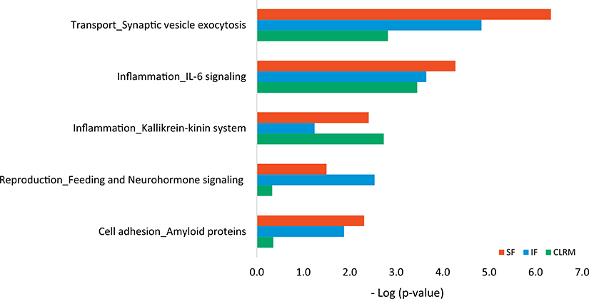
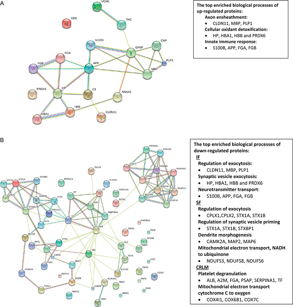
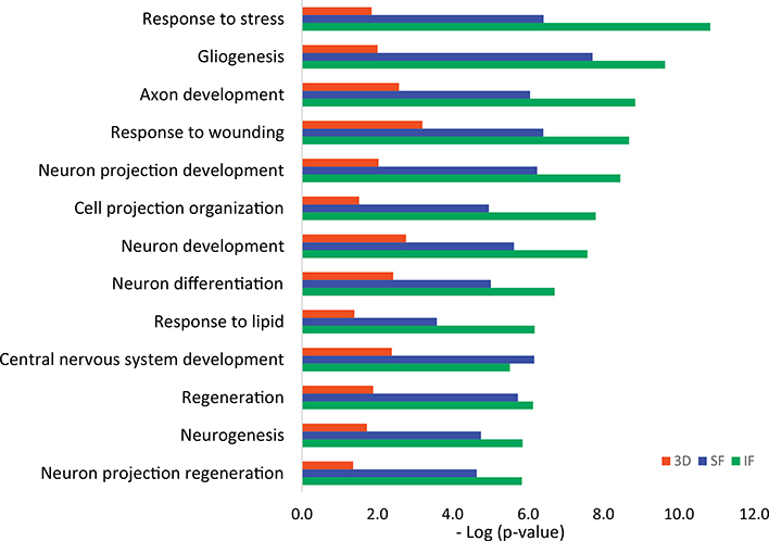
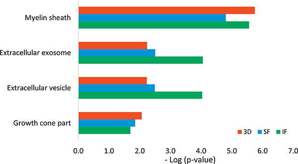
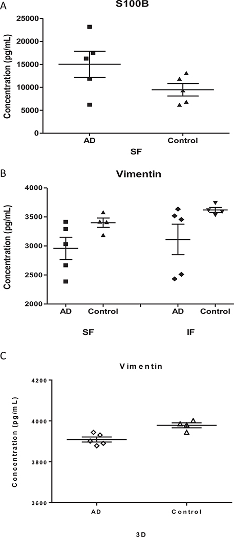
We established a unique platform for proteomic analysis of cultured three-dimensional (3D) neurons and brain tissue from Alzheimer's disease (AD) patients. We collected peripheral blood mononuclear cells (PBMC), converted PBMC to induced pluripotent stem cell (iPSC) lines, and differentiated the iPSC into human 3D neuro-spheroids. The postmortem brain tissue from the superior frontal cortex, inferior frontal cortex and cerebellum area of the AD patients was compared to the same regions from the control subjects. Proteomic analysis of 3D neuro-spheroids derived from AD subjects revealed the alteration of a number of proteins involved in axon growth, mitochondrial function, and antioxidant defense. Similar analysis of post-mortem AD brain tissue revealed significant alteration in proteins involved in oxidative stress, neuro-inflammation, along with proteins related to axonal injury. These results clearly indicate that the dysfunction of 3D neurons from AD patients in our in vitro environment is comparable to the post-mortem AD brain tissue in vivo. In conclusion, our study revealed a number of candidate proteins that have important implications in AD pathogenesis and supports the notion that the iPSC-derived 3D neuronal system functions as a model to examine novel aspects of AD pathology.
Significance: In this study, we present a unique platform for proteomic analysis of induced pluripotent stem cell-derived three dimensional (3D) neurons and compare the results to those from three regions of post-mortem brain tissue from Alzheimer's disease patients and normal control subjects. Our results show that the dysfunction of 3D neurons from AD patients in our in vitro environment is comparable to the post-mortem AD brain tissue in vivo. Our results revealed several candidate proteins that have important implications in AD pathogenesis.
Keywords: 3D; Alzheimer; Bioinformatic; Neuro-spheroid; Proteomics; iPSC.
Copyright © 2018. Published by Elsevier B.V.
Figures
References
-
- Burns A, Iliffe S, Alzheimer’s disease, BMJ 338 (2009) b158. - PubMed
-
- Tanzi RE, Bertram L, Twenty years of the Alzheimer’s disease amyloid hypothesis: a genetic perspective, Cell 120 (2005) 545–555. - PubMed
-
- Querfurth HW, LaFerla FM, Alzheimer’s disease, N. Engl. J. Med 362 (2010) 329–344. - PubMed
-
- Selkoe DJ, Alzheimer’s disease is a synaptic failure, Science 298 (2002) 789–791. - PubMed
-
- van der Zee J, Sleegers K, Van Broeckhoven C, Invited article: the Alzheimer disease-frontotemporal lobar degeneration spectrum, Neurology 71 (2008) 1191–1197. - PubMed
Publication types
MeSH terms
Substances
Grants and funding
LinkOut - more resources
Full Text Sources
Other Literature Sources
Medical
