Review
. 2018 Apr 28;15(5):878.
doi: 10.3390/ijerph15050878.

Exercise Early and Often: Effects of Physical Activity and Exercise on Women's Bone Health

Affiliations
Review

Exercise Early and Often: Effects of Physical Activity and Exercise on Women's Bone Health

Karen L Troy et al. Int J Environ Res Public Health. .

Abstract

In 2011 over 1.7 million people were hospitalized because of a fragility fracture, and direct costs associated with osteoporosis treatment exceeded 70 billion dollars in the United States. Failure to reach and maintain optimal peak bone mass during adulthood is a critical factor in determining fragility fracture risk later in life. Physical activity is a widely accessible, low cost, and highly modifiable contributor to bone health. Exercise is especially effective during adolescence, a time period when nearly 50% of peak adult bone mass is gained. Here, we review the evidence linking exercise and physical activity to bone health in women. Bone structure and quality will be discussed, especially in the context of clinical diagnosis of osteoporosis. We review the mechanisms governing bone metabolism in the context of physical activity and exercise. Questions such as, when during life is exercise most effective, and what specific types of exercises improve bone health, are addressed. Finally, we discuss some emerging areas of research on this topic, and summarize areas of need and opportunity.

Keywords: areal bone mineral density; bone adaptation; bone mineral density; high resolution peripheral quantitative computed tomography; mechanical loading; quantitative computed tomography; structure; volumetric bone mineral density.


Conflict of interest statement

The authors declare no conflicts of interest.

Figures

Figure 1
Figure 1
Current available methods for the assessment of bone strength and fracture risk. (A) Dual energy X-ray absorptiometry (DXA) forearm scan with standard ultradistal (UD), middle (MID) and one-third of arm length (1/3) regions, used to calculate aBMD (g/cm2). (B) 3D view of clinical computed tomography (CT) scan of the distal radius, with (C) coronal view containing dotted line indicating position of (D) transverse view. CT scan acquired at a transverse pixel size of 234 μm and slice thickness of 625 μm. (E) 3D view of high resolution peripheral quantitative CT (HRpQCT) image (F) of the distal radius, with isotropic voxel size of 82 μm.
Figure 2
Figure 2
Typical pattern of age-related changes in bone mass, which is primarily accrued during the pre-pubertal and adolescent stages, reaches a lifetime peak at approximately 18 years of age, and declines sharply during perimenopause and steadily post-menopause.
Figure 3
Figure 3
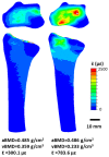
Bone strain (expressed as energy-equivalent strain, ε¯ [24]) in the distal radius and transverse slice with maximum cross-sectional area. Percent difference in aBMD is 0.21%, while percent difference in vBMD and mean energy equivalent strain in the 9.375 mm ultradistal region is are 42.68% and 89.23%, respectively.
Figure 4
Figure 4
Distal radius microstructure acquired using HR-pQCT viewed from the transverse plane (right) and sagittal cross-section (top left). Insets show example measurements of compartment-specific cortical (porosity) and trabecular (number, thickness) microstructure parameters, made possible through this emerging technology.

References

    1. United States Bone and Joint Initiative . In: The Burden of Musculoskeletal Diseases in the United States. 3rd ed. Saag K., Wright N.C., Kilgore M., editors. American Academy or Orthopaedic Surgeons; Rosemont, IL, USA: 2014.
    1. Kanis J.A., Oden A., Johansson H., Borgström F., Ström O., McCloskey E. FRAX®and its applications to clinical practice. Bone. 2009;44:734–743. doi: 10.1016/j.bone.2009.01.373. - DOI - PubMed
    1. Hernandez C.J., Beaupré G.S., Carter D.R. A theoretical analysis of the relative influences of peak BMD, age-related bone loss and menopause on the development of osteoporosis. Osteoporos. Int. 2003;14:843–847. doi: 10.1007/s00198-003-1454-8. - DOI - PubMed
    1. Cosman F., Lindsay R., LeBoff M.S., Jan de Beur S., Tanner B. Clinician’s Guide to Prevention and Treatment of Osteoporosis. Natl. Osteoporos. Found. 2014;1:55. doi: 10.1007/s00198-014-2794-2. - DOI - PMC - PubMed
    1. National Osteoporosis Foundation Osteoporosis Exercise for Strong Bones. [(accessed on 3 March 2018)]; Available online: https://www.nof.org/patients/fracturesfall-prevention/exercisesafe-movem...

Publication types