. 2018 May 15;115(20):E4661-E4669.
doi: 10.1073/pnas.1720065115. Epub 2018 Apr 30.

Distinct roles of resident and nonresident macrophages in nonischemic cardiomyopathy

Affiliations

Distinct roles of resident and nonresident macrophages in nonischemic cardiomyopathy

Xudong Liao et al. Proc Natl Acad Sci U S A. .

Abstract

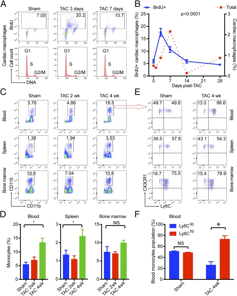
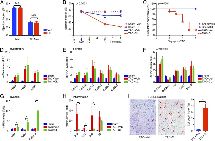
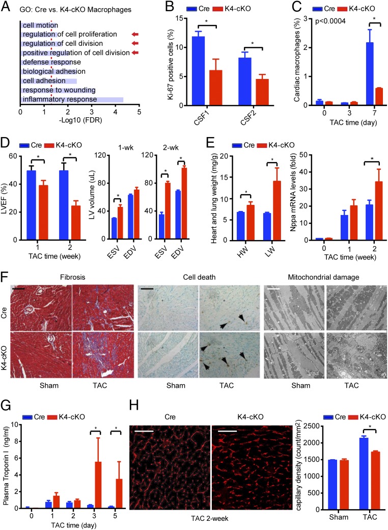
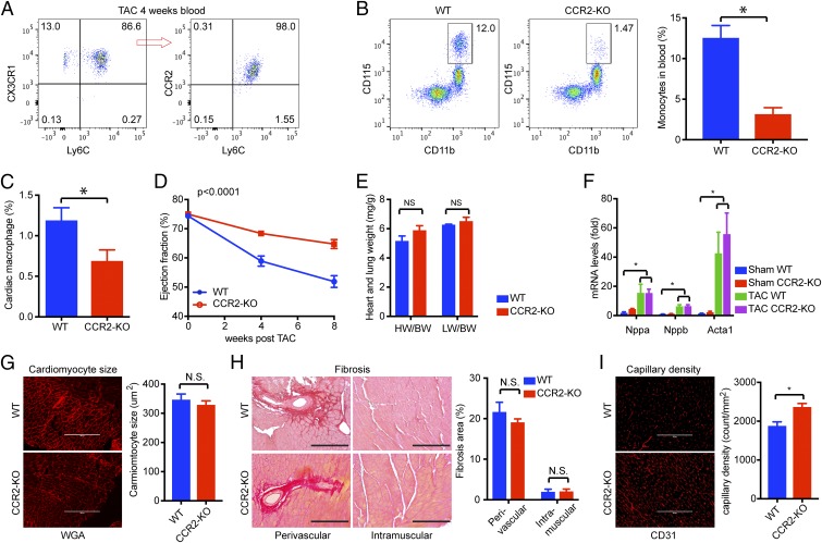
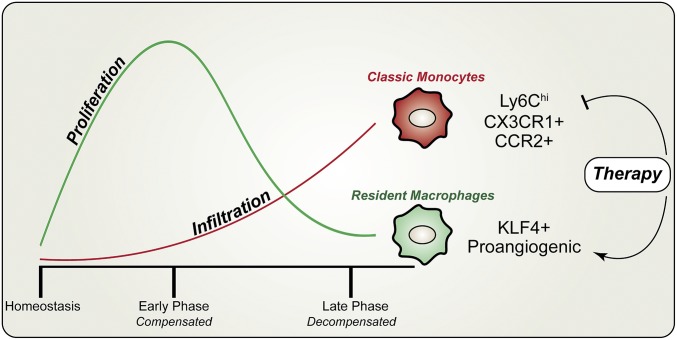
Nonischemic cardiomyopathy (NICM) resulting from long-standing hypertension, valvular disease, and genetic mutations is a major cause of heart failure worldwide. Recent observations suggest that myeloid cells can impact cardiac function, but the role of tissue-intrinsic vs. tissue-extrinsic myeloid cells in NICM remains poorly understood. Here, we show that cardiac resident macrophage proliferation occurs within the first week following pressure overload hypertrophy (POH; a model of heart failure) and is requisite for the heart's adaptive response. Mechanistically, we identify Kruppel-like factor 4 (KLF4) as a key transcription factor that regulates cardiac resident macrophage proliferation and angiogenic activities. Finally, we show that blood-borne macrophages recruited in late-phase POH are detrimental, and that blockade of their infiltration improves myocardial angiogenesis and preserves cardiac function. These observations demonstrate previously unappreciated temporal and spatial roles for resident and nonresident macrophages in the development of heart failure.

Keywords: angiogenesis; cardiac macrophage; pressure overload hypertrophy.


Conflict of interest statement

Conflict of interest statement: X.L. and M.K.J. have a patent application (serial no. 62/644,792).

Figures

Fig. 1.
Fig. 1.
Pressure overload induces local proliferation of cardiac resident macrophages at early phase and infiltration of monocytes at late phase. (A) Gating strategy for flow cytometry. (B) Dynamics of cardiac macrophages following TAC and its correlation with cardiac function. The dashed line indicates baseline. Cardiac macrophages were gated as live (live/dead stain-negative) CD45+CD11b+Ly6GF4/80+CD64+ cells and are shown as a percentage vs. total heart cells. Cardiac function was expressed as left ventricular fractional shortening. (C) Immunostaining for Mac-3 to reveal macrophages in the myocardium, particularly in the perivascular region. Brown (DAB-positive) cells are macrophages. Arrowheads indicate vessels. (Scale bars, 100 μm.) (D) LV dysfunction following TAC. (E) Strategy to generate CD45.1-CD45.2 chimeric mice. Recipient CD45.2 mice were irritated with their head-chest shielded by lead block and transplanted via tail vein injection with bone marrow cells from CD45.1 donor mice. The resulting chimera mice carry both CD45.2 and CD45.1 cells in the blood but only have CD45.2 cardiac resident macrophages. (F) Blood (50 μL) was drawn from the tail vein at 8 wk posttransplantation and analyzed by FACS for CD45.1+ myeloid cells (CD45+CD11b+). (G) FACS analysis for CD45.1+ cardiac macrophages before and after TAC. (H) Corrected monocyte infiltration rate considering that only 50% of monocytes were CD45.1+ cells. (I) FACS analysis for Ki-67 expression in cardiac macrophages. (B and D) P values were calculated from one-way ANOVA. (FI) *P < 0.05 by t test after Bonferroni correction (n = 5–8 for all data points). NS, not significant.
Fig. 2.
Fig. 2.
Local proliferation of cardiac resident macrophages and infiltration of blood-borne Ly6C+ monocytes. (A and B) TAC induced local proliferation of cardiac resident macrophages as assessed by BrdU pulse-labeling assay. DNA was stained with Hoechst 33342 for cycle analysis, and the S-phase was clearly visible in TAC 3-d samples. The time course curve of total cardiac macrophages (dotted red line) was overlaid to show the phase difference. (C and D) Monocytes (CD115+/CD11b+) in blood, spleen, and bone marrow before and after TAC. (E and F) Classic Ly6C+ monocytes and nonclassic Ly6C monocytes before and after TAC. In B, P values were calculated from one-way ANOVA for BrdU data. In D and F, *P < 0.05 by t test (n = 5 for all data points). NS, not significant.
Fig. 3.
Fig. 3.
Cardiac resident macrophages are requisite for cardiac adaptation to POH. (A) Blocking monocyte infiltration by RS had no effect on POH. (B) Left ventricular function following clodronate-mediated macrophage depletion and TAC (n = 8–10 in each group). The P value was calculated from two-way ANOVA. The blue arrow indicates the time of TAC, and red arrows indicate the time of liposome injections. (C) Postoperation survival rates. P values were calculated from log-rank between TAC+CL and TAC+Veh groups. (DH) Gene expression in the heart after 4-d TAC ± CL (n = 5). *P < 0.05 by t test with Bonferroni correction. (I) Cell death detected by TUNEL staining in the myocardium after 4-d TAC ± CL. Red arrowheads indicate nuclei of dead cells. *P < 0.05 by t test (n = 5). NS, not significant. (Scale bars, 200 μm.)
Fig. 4.
Fig. 4.
Myeloid KLF4 regulates cardiac macrophage proliferation in POH. (A) RNA-seq studies with primary peritoneal macrophages from Cre and K4-cKO mice. A total of 283 genes were differentially regulated between the Cre and K4-cKO groups, and gene ontology (GO) term enrichment analysis revealed nine pathways that were functionally enriched [false discovery rate (FDR) < 0.05]. Arrows indicate pathways of interest. (B) KLF4-deficient peritoneal macrophages exhibited an impaired proliferative response to CSF1/2 stimulation (48 h). *P < 0.05. (C) KLF4-deficiency impaired TAC-induced cardiac macrophage proliferation (n = 10). *P < 0.05. (D) Myeloid KLF4 deficiency impaired cardiac function after TAC, resulting in dilated cardiomyopathy at 2 wk post-TAC (n = 8–10). *P < 0.05. EDV, end-diastolic volume; ESV, end-systolic volume; LVEF, left ventricular ejection fraction. (E) Myeloid KLF4 deficiency accelerated TAC-induced cardiac hypertrophy. Heart weight (HW; milligrams) and lung weight (LW; milligrams) at 2 wk post-TAC were normalized to body weight (grams) (n = 6 in each group). *P < 0.05 by t test with Bonferroni correction. (F) TAC induced more fibrosis, cell death, and mitochondrial damage in K4-cKO hearts. Samples were from 1-wk TAC and sham mice. (Black scale bar, 100 μm; white scale bar, 2 μm.) (G) TAC induced higher plasma cardiac troponin I levels in the K4-cKO group (n = 5). *P < 0.05 with Bonferroni correction. (H) Impaired angiogenesis in K4-cKO hearts after TAC as revealed by myocardial capillary density using CD31 immunostaining (n = 5). *P < 0.05. (Scale bars, 100 μm.).
Fig. 5.
Fig. 5.
Blockade-infiltrating macrophages preserved cardiac function in late-phase POH. (A) Classic Ly6C+CCR2+ monocytes increased in blood after 4 wk of TAC. A representative FACS plot from five samples is shown. (B) Reduced monocyte numbers in CCR2-KO mice after 4 wk of TAC (n = 5). *P < 0.05 by t test. (C) Reduced cardiac macrophage numbers in CCR2-KO hearts after 4 wk of TAC (n = 5). *P < 0.05 by t test. (D) Left ventricular function assessed by echocardiography. The P value is shown as P (genotype * time) calculated from two-way ANOVA. (E) Heart weight and lung weight. (F) Expression of hypertrophy marker genes. (G) Cardiomyocyte cross-section area. Cell membranes were outlined by wheat germ agglutinin (WGA) staining. (H) Fibrosis assessed by PicoSirus Red staining, with collagen stained in red. Perivascular and intramuscular areas are shown. Fibrosis was quantified as the percentage of the fibrotic area in total by ImageJ (NIH) software. (I) Myocardial capillary density assessed by CD31 staining. All tissue samples were assessed at 8 wk post-TAC (n = 5–8). *P < 0.05. NS, not significant. (Scale bars, 200 μm.)
Fig. 6.
Fig. 6.
Dynamic changes and distinct roles of cardiac macrophage subsets in POH. Our studies have demonstrated a two-phase response of cardiac macrophages to pressure overload. (i) During the early phase of POH, there is robust local proliferation of resident macrophages that is crucial for cardiac compensation to pressure overload. (ii) During the late phase of POH, the infiltration of inflammatory monocytes increases, which promotes the transition to cardiac decompensation. Mechanistically, transcription factor KLF4 is required for resident macrophage proliferation and the infiltrating monocytes are Ly6ChiCX3CR1+CCR2+ classic monocytes. Finally, this model implies that these two subsets of cardiac macrophages can be targeted for therapeutic gain, either by blocking monocytes (i.e., RS administration) or by promoting resident macrophages (i.e., prolonged proliferation), or by simultaneously targeting both subsets for a healthy balance.

References

    1. Cecchi F, et al. Coronary microvascular dysfunction and prognosis in hypertrophic cardiomyopathy. N Engl J Med. 2003;349:1027–1035. - PubMed
    1. Treasure CB, et al. Hypertension and left ventricular hypertrophy are associated with impaired endothelium-mediated relaxation in human coronary resistance vessels. Circulation. 1993;87:86–93. - PubMed
    1. Maron BJ, Wolfson JK, Epstein SE, Roberts WC. Intramural (“small vessel”) coronary artery disease in hypertrophic cardiomyopathy. J Am Coll Cardiol. 1986;8:545–557. - PubMed
    1. Frey N, Luedde M, Katus HA. Mechanisms of disease: Hypertrophic cardiomyopathy. Nat Rev Cardiol. 2011;9:91–100. - PubMed
    1. Sano M, et al. p53-induced inhibition of Hif-1 causes cardiac dysfunction during pressure overload. Nature. 2007;446:444–448. - PubMed

Publication types

MeSH terms

Substances