. 2018 Apr 30;9(1):1715.
doi: 10.1038/s41467-018-04194-9.

Piezo1 is a mechanically activated ion channel and mediates pressure induced pancreatitis

Affiliations

Piezo1 is a mechanically activated ion channel and mediates pressure induced pancreatitis

Joelle M-J Romac et al. Nat Commun. .

Abstract

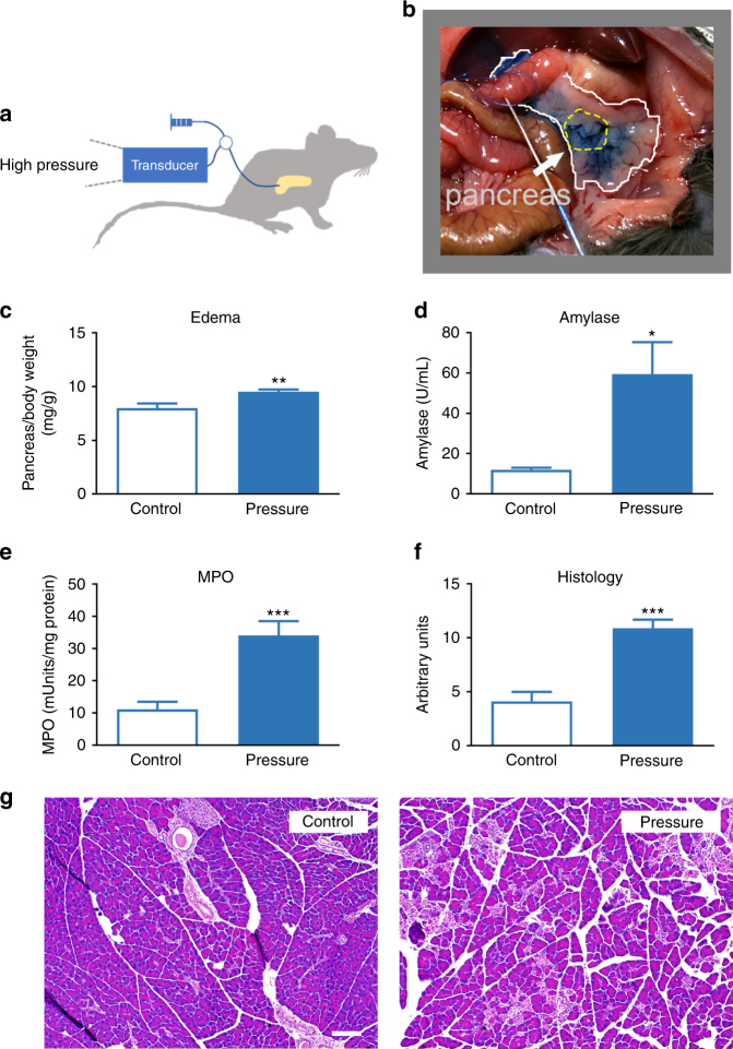
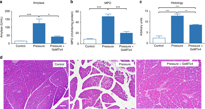
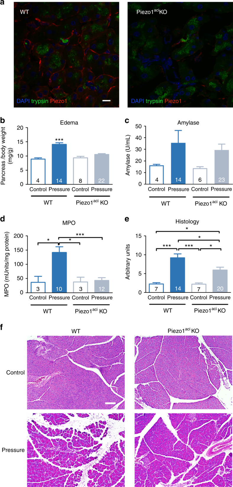
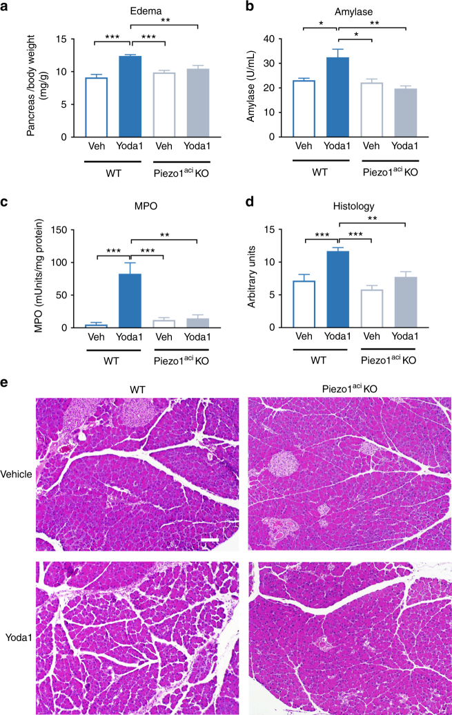
Merely touching the pancreas can lead to premature zymogen activation and pancreatitis but the mechanism is not completely understood. Here we demonstrate that pancreatic acinar cells express the mechanoreceptor Piezo1 and application of pressure within the gland produces pancreatitis. To determine if this effect is through Piezo1 activation, we induce pancreatitis by intrapancreatic duct instillation of the Piezo1 agonist Yoda1. Pancreatitis induced by pressure within the gland is prevented by a Piezo1 antagonist. In pancreatic acinar cells, Yoda1 stimulates calcium influx and induces calcium-dependent pancreatic injury. Finally, selective acinar cell-specific genetic deletion of Piezo1 protects mice against pressure-induced pancreatitis. Thus, activation of Piezo1 in pancreatic acinar cells is a mechanism for pancreatitis and may explain why pancreatitis develops following pressure on the gland as in abdominal trauma, pancreatic duct obstruction, pancreatography, or pancreatic surgery. Piezo1 blockade may prevent pancreatitis when manipulation of the gland is anticipated.


Conflict of interest statement

The authors declare no competing interests.

Figures

Fig. 1
Fig. 1
Intrapancreatic duct pressure causes acute pancreatitis. a Test solutions were infused into the pancreatic duct of mice as illustrated. Injection pressures were continuously monitored using a pressure transducer. b Injection of 400 µL buffered saline containing methylene blue dye was distributed throughout the pancreas. The white line indicates the boundary of the pancreas and yellow line indicates the mid-region of the pancreas that was used for histology and myeloperoxidase (MPO) determinations. Either sham-operated animals (control) or buffered saline solutions were infused at 25–33 mm Hg (pressure) and c edema, d blood amylase, e MPO, and f histological scoring were analyzed 24 h after duct injection in C57BL/6J male mice. Statistical analysis was performed using Student’s t test. *P ≤ 0.05; **P ≤ 0.01; ***P ≤ 0.001 (n = 7–13 for control and 13–17 for pressure). g Representative images of pancreatic tissue stained with H&E. Bar = 100 µm
Fig. 2
Fig. 2
Piezo1 is expressed in pancreatic acinar cells. a The relative expression of mechanically activated ion channel messenger RNAs from C57BL/6J pancreatic acinar cells was analyzed by real-time RT-PCR using the ΔΔCT method (biological n = 3). b Frozen sections (left panel) from mouse pancreatic tissue (C57BL/6J) were immunostained with antibodies against trypsin 3 (green) or Piezo1 (red). DAPI-stained nuclei are shown in blue. Inset shows a higher magnification of an acinar cell. Images were taken with a ×63 objective. Bar = 10 µm. c Representative live-cell imaging of pancreatic acini loaded with Calcium 6-QF at time 0 (left panel) and at the time of maximum fluorescence (right panel). Images obtained with a ×20 objective. Bar = 10 µm. d A representative time course of calcium fluorescence in acinar cells from littermate wild-type or Piezo1aci KO mice. Black arrow indicates when Yoda1 (25 µM) was added to the cells. e Statistical analysis of Fmax/F0 in acinar cells from littermate wild-type or Piezo1aci KO mice. GsMTx4 was added 2 min prior to Yoda1 (n = 3–5 experiments; 30–40 cells were analyzed for each experiment). LDH released from acini treated with Yoda1 (100 μM) from f C57BL/6J mice or g Piezo1aci KO and littermate (WT) mice (n = 4–5). Results are expressed as fold changes compared to the control condition (absence of test agents). DMSO was the vehicle for Yoda1; fold changes were determined vs. LDH release from cells that were incubated in absence of vehicle or compound. Statistical analysis was performed using ANOVA test with Tukey’s post-test. *P ≤ 0.05; **P ≤ 0.01; ***P ≤ 0.001
Fig. 3
Fig. 3
The Piezo1 antagonist GsMTx4 reduces the severity of pancreatitis. C57BL/6J mice were either sham-operated (control) or received intrapancreatic duct infusion of buffered saline at 25–33 mm Hg (pressure). GsMTx4 (270 µg/kg) was administered by intraperitoneal injection 1 h prior to intrapancreatic duct infusion of saline at 25–33 mm Hg (pressure + GsMTx4). a Blood amylase, b pancreatic MPO, and c histological scoring of pancreatitis severity were measured. Statistical analysis was performed using ANOVA test with Tukey’s post-test. *P ≤ 0.05; **P ≤ 0.01; ***P ≤ 0.001 (n = 6). d Representative images of pancreatic tissue stained with H&E. Bar = 100 µm
Fig. 4
Fig. 4
Genetic deletion of Piezo1 in pancreatic acinar cells reduces the severity of pancreatitis. a Confocal microscopic images of pancreatic sections from littermate wild-type (left panel) and Piezo1aci KO (right panel) mice imunostained for Piezo1 (red) and trypsin (green). DAPI-stained nuclei are blue. ×63 objective. Bar = 10 µm. Buffered saline was injected into the pancreatic duct at 25–33 mm Hg (pressure) of Piezoaci KO or littermate wild-type (WT) mice. Control animals were subjected to sham operation. Tissues were harvested 24 h post injection and analyzed for b pancreatic edema, c blood amylase, d MPO, and e histological scoring. The number of animals in each group is indicated in the respective column. Statistical analysis was performed using ANOVA test with Tukey’s post-test. *P ≤ 0.05; ***P ≤ 0.001. f Representative images of pancreatic tissue stained with H&E taken with a ×10 objective. Bar = 100 µm
Fig. 5
Fig. 5
The Piezo1 agonist Yoda1 induces acute pancreatitis in wild-type mice but not in mice with genetic deletion of Piezo1. Yoda1 at a dose of 0.4 mg/kg in 50 µL (1.1 % DMSO; 4.8% ethanol; 94.1% buffered saline, pH 7.6) was infused into the pancreatic duct of WT or Piezo1aci KO mice over 10 min at pressures not exceeding 7–11 mm Hg. Either vehicle or Yoda1 was infused. Tissues were harvested 24 h post injection and analyzed for a pancreatic edema, b serum amylase, c pancreatic myeloperoxidase (MPO), and d histological scoring. n = 6–8 animals per group. Statistical analysis was performed by ANOVA with Tukey’s post-test. *P ≤ 0.05; **P ≤ 0.01; ***P ≤ 0.001. e Representative images of pancreatic tissue stained with H&E are shown. Bar = 100 µm

References

    1. Malgras B, Douard R, Siauve N, Wind P. Management of left pancreatic trauma. Am. Surg. 2011;77:1–9. - PubMed
    1. Opie EL, Meakins JC. Data concerning the etiology and pathology of hemorrhagic necrosis of the pancreas (acute hemorrhagic pancreatitis) J. Exp. Med. 1909;11:561–578. doi: 10.1084/jem.11.4.561. - DOI - PMC - PubMed
    1. Lerch MM, et al. Pancreatic duct obstruction triggers acute necrotizing pancreatitis in the opossum. Gastroenterology. 1993;104:853–861. doi: 10.1016/0016-5085(93)91022-A. - DOI - PubMed
    1. Wang AY, Strand DS, Shami VM. Prevention of post-endoscopic retrograde cholangiopancreatography pancreatitis: medications and techniques. Clin. Gastroenterol. Hepatol. 2016;14:1521–1532. doi: 10.1016/j.cgh.2016.05.026. - DOI - PubMed
    1. Freeman ML, Guda NM. Prevention of post-ERCP pancreatitis: a comprehensive review. Gastrointest. Endosc. 2004;59:845–864. doi: 10.1016/S0016-5107(04)00353-0. - DOI - PubMed

Publication types

MeSH terms