. 2018 Jan:50:459-482.
doi: 10.1146/annurev-fluid-122316-045259.

Lymphatic System Flows

Affiliations

Lymphatic System Flows

James E Moore Jr et al. Annu Rev Fluid Mech. 2018 Jan.

Abstract

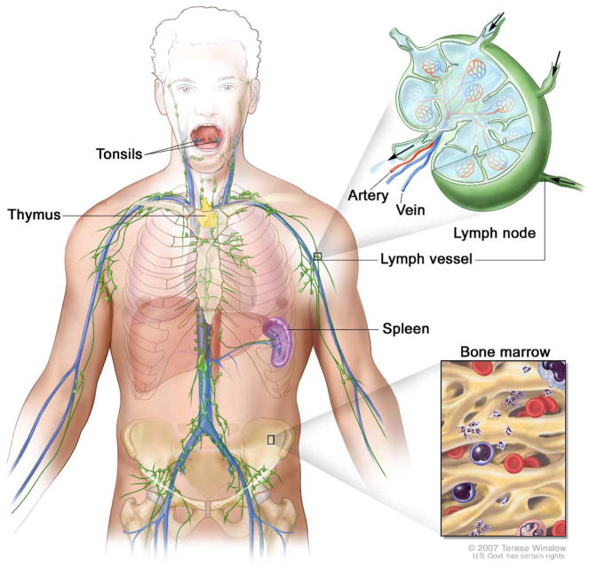
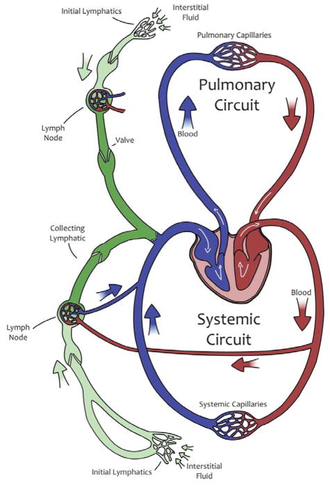
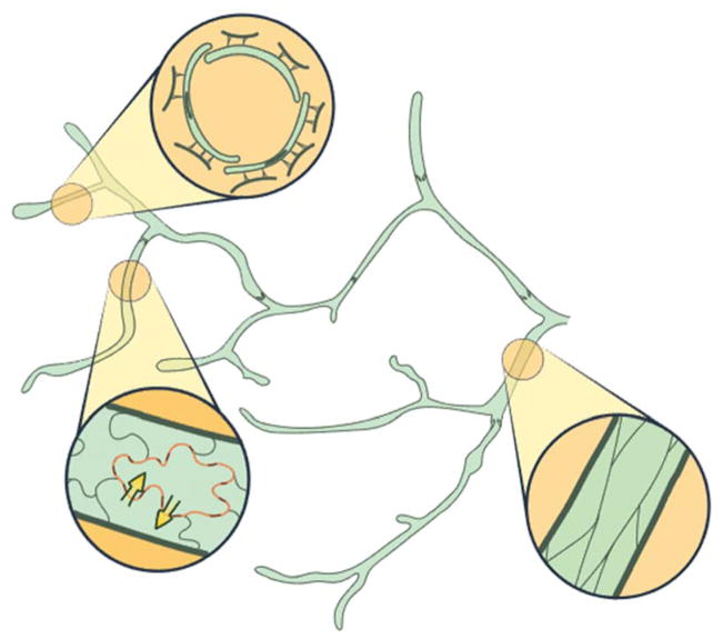
The supply of oxygen and nutrients to tissues is performed by the blood system, and involves a net leakage of fluid outward at the capillary level. One of the principal functions of the lymphatic system is to gather this fluid and return it to the blood system to maintain overall fluid balance. Fluid in the interstitial spaces is often at subatmospheric pressure, and the return points into the venous system are at pressures of approximately 20 cmH2O. This adverse pressure difference is overcome by the active pumping of collecting lymphatic vessels, which feature closely spaced one-way valves and contractile muscle cells in their walls. Passive vessel squeezing causes further pumping. The dynamics of lymphatic pumping have been investigated experimentally and mathematically, revealing complex behaviours indicating that the system performance is robust against minor perturbations in pressure and flow. More serious disruptions can lead to incurable swelling of tissues called lymphœdema.

Keywords: Cancer; Immunology; Lymph; Oedema; Physiology.


Figures

Figure 1
Figure 1
The organs of the lymphatic system. Major lymph vessels in the trunk and upper limbs are shown in green (Institute).
Figure 2
Figure 2
Schematic of the blood circulation and lymphatic vascular system. Some lymph fluid is reabsorbed via the nodal blood circulation under normal conditions, resulting in post-nodal lymph having a higher protein concentration. Based on (Lubopitko).
Figure 3
Figure 3
A small network of initial lymphatics. Top inset shows endothelial-cell primary valves, consisting of unbonded overlaps between ECs, and anchoring filaments to surrounding fibrous tissue. Lower left inset shows characteristic oak-leaf EC configuration, with discontinuous button junctions. Little is known about where, i.e. how far along the network, such cells give way to ECs with continuous zipper junctions (inset at bottom right). Although not divided into lymphangions, initial lymphatics can also have sparse secondary (intravascular) valves. Based on published material (Baluk et al 2007, Galie & Spilker 2009, Murfee et al 2007, Schmid-Schönbein 1990a).
Figure 4
Figure 4
Subcutaneous lymph capillaries and deeper-lying collecting lymphatics in the leg of a 130mm human fœtus. From (Kampmeier 1928). Scale: long edge = 4 mm approx.
Figure 5
Figure 5
A transverse section of a lymphatic valve from rat mesentery, showing the two leaflets, the sinus, and the lymphatic vessel continuing at each end. Visualisation by fluorescently tagged nitric oxide synthase expressed by the lymphatic endothelial cells. From (Bohlen et al 2009).
Figure 6
Figure 6
Two orientations of a 3D reconstruction of a lymphatic valve from a stack of confocal images. Left, looking downstream, into the narrowing bore of the valve between the leaflets, and at the outside of the vessel wall as it tapers outwards to the middle of the sinus. Right: looking upstream, directly at the free trailing edge of the leaflets and the twin blind fluid sacs formed between the inside wall of the sinus and the outer surface of the leaflets. From (Zawieja 2009).
Figure 7
Figure 7
Comparison of the passive pressure/diameter relation of a lymphatic, a venule and an arteriole, all from the mesentery of the same rat. Diameter is normalised to the maximum value in each case: lymphatic 267 μm, venule 278 μm, and arteriole 135 μm. The non-linearity of elastic stiffness is related to the increase in local slope between (say) Dnorm = 0.8 and Dnorm = 1. The increase at high distending pressure is much more for the venule and lymphatic than the arteriole, and (as shown in the inset) more for the lymphatic than the venule. Redrawn from (Rahbar et al 2012).
Figure 8
Figure 8
Lumped-parameter modelling simulations of pumping by a chain of lymphangions contracting synchronously in the presence of positive (left) and negative (right) transmural pressure. D = diameter of the first lymphangion (indicated by star, at top), p = intra-lymphangion pressure, Q = flow-rate through the first valve in the chain, M = contraction activation, Δptm = transmural pressure, t = time. Pumping is initiated as shown in the curve of M(t) by lymphatic muscle activation (red), followed by relaxation (green) and inactivation (yellow). These colours are then used in the other panels to indicate timing. Suction occurs just after 170 seconds, when the pressure inside the lymphangion dips below the inlet pressure pa, after which the flow-rate through the first valve peaks. The loops of Δptm vs. D illustrate the time-course alongside the passive behaviour of the lymphangion (black), with the area of the loop defining the output work, and change in diameter indicating the flow-rate generated. In the presence of a negative transmural pressure (right), the Δptm-D loop is so small as to be barely visible, indicating pumping failure. Animations available in the on-line version.

Similar articles

Cited by

References

    1. Adair TH, Guyton AC. Modification of lymph by lymph nodes. II. Effect of increased lymph node venous blood pressure. American Journal of Physiology (Heart Circ Physiol ) 1983;245:H616–H22. - PubMed
    1. Adair TH, Guyton AC. Modification of lymph by lymph nodes III. Effect of increased lymph hydrostatic pressure. American Journal of Physiology (Heart Circ Physiol ) 1985;249:H777–H82. - PubMed
    1. Allanson JE. Lymphatic System. In: Stevenson RE, Hall JG, editors. Human Malformations and Related Anomalies. 2. New York: Oxford University Press; 2005. pp. 145–82.
    1. Aukland K, Reed RK. Interstitial-lymphatic mechanisms in the control of extracellular fluid volume. Physiological Reviews. 1993;73:1–78. - PubMed
    1. Baish JW, Kunert C, Padera TP, Munn LL. Synchronization and random triggering of lymphatic vessel contractions. PLoS Computational Biology. 2016;12:e1005231. - PMC - PubMed

LinkOut - more resources