

Ultrasonographic Findings of Scleredema Adultorum of Buschke Involving the Posterior Neck
- PMID: 29713220
- PMCID: PMC5904469
- DOI: 10.3348/kjr.2018.19.3.425
Ultrasonographic Findings of Scleredema Adultorum of Buschke Involving the Posterior Neck
Abstract
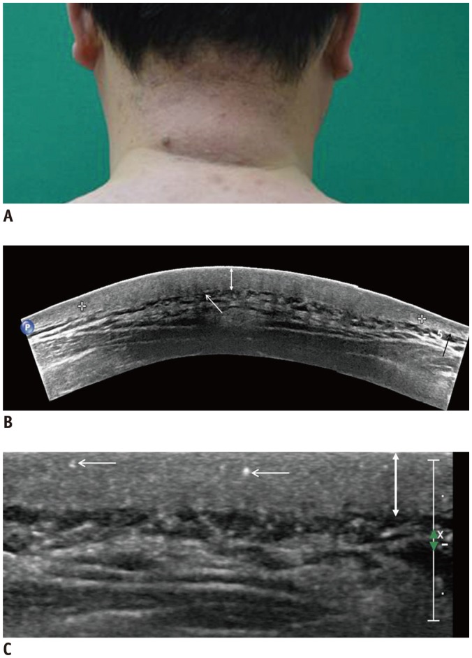
Objective: To describe the clinical and ultrasonographic (US) findings in patients with scleredema adultorum of Buschke, who presented with sclerotic skin on their posterior neck.
Materials and methods: After obtaining IRB approval, eight patients with scleredema adultorum of Buschke were enrolled. They underwent US examination of their posterior neck. The diagnoses were confirmed pathologically. The clinical history and US images were evaluated retrospectively. Dermal thickness was compared between the patient group and the age- and sex-matched control group.
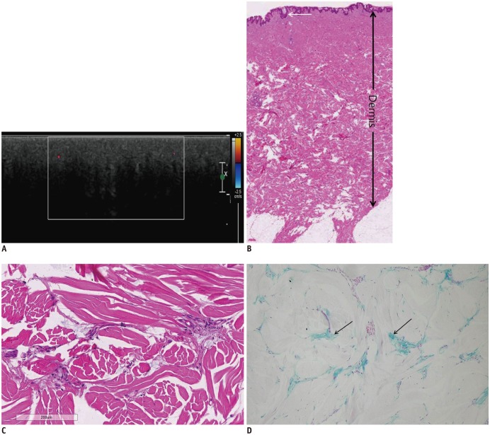
Results: The patients included seven males and one female with a mean age of 51.5 years. All patients presented with thickening of the skin and/or a palpable mass on the posterior neck. Five (62.5%) of the eight patients showed erythematous discoloration. Six patients (75.0%) had a history of diabetes. The Hemoglobin A1c level was found to be increased in all patients. US images did not show any evidence of a soft tissue mass or infection. The mean dermal thickness in patients (7.01 ± 1.95 mm) was significantly greater than that in the control group (3.08 ± 0.87 mm) (p = 0.001). Multiple strong echogenic spots in the dermis were seen in all patients. Seven patients (87.5%) showed posterior shadowing in the lower dermis.
Conclusion: When a patient with a history of diabetes presents with a palpable mass or erythematous discoloration of the posterior neck and US shows the following imaging features: 1) no evidence of a soft tissue mass or infection, 2) thickening of the dermis, 3) multiple strong echogenic spots and/or posterior shadowing in the dermis, scleredema adultorum of Buschke should be considered in the differential diagnosis.
Keywords: Dermis; Diabetes; Neck; Scleredema; Soft tissue; Ultrasonography.
Figures
References
-
- Foti R, Leonardi R, Rondinone R, Di Gangi M, Leonetti C, Canova M, et al. Scleredema-like disorders. Autoimmun Rev. 2008;7:331–339. - PubMed
-
- Fabri M, Hunzelmann N. [Differential diagnosis of scleredema and pseudoscleredema] J Dtsch Dermatol Ges. 2007;5:977–984. - PubMed
-
- Meguerditchian C, Jacquet P, Béliard S, Benderitter T, Valéro R, Carsuzza F, et al. Scleredema adultorum of Buschke: an under recognized skin complication of diabetes. Diabetes Metab. 2006;32(5 Pt 1):481–484. - PubMed
-
- Rho YW, Suhr KB, Lee JH, Park JK. A clinical observation of scleredema adultorum and its relationship to diabetes. J Dermatol. 1998;25:103–107. - PubMed
-
- Ioannidou DI, Krasagakis K, Stefanidou MP, Karampekios S, Panayiotidis J, Tosca AD. Scleredema adultorum of Buschke presenting as periorbital edema: a diagnostic challenge. J Am Acad Dermatol. 2005;52(2 Suppl 1):41–44. - PubMed
Publication types
MeSH terms
Substances
LinkOut - more resources
Full Text Sources
Other Literature Sources
