

Nodule Classification on Low-Dose Unenhanced CT and Standard-Dose Enhanced CT: Inter-Protocol Agreement and Analysis of Interchangeability
- PMID: 29713230
- PMCID: PMC5904479
- DOI: 10.3348/kjr.2018.19.3.516
Nodule Classification on Low-Dose Unenhanced CT and Standard-Dose Enhanced CT: Inter-Protocol Agreement and Analysis of Interchangeability
Abstract
Objective: To measure inter-protocol agreement and analyze interchangeability on nodule classification between low-dose unenhanced CT and standard-dose enhanced CT.
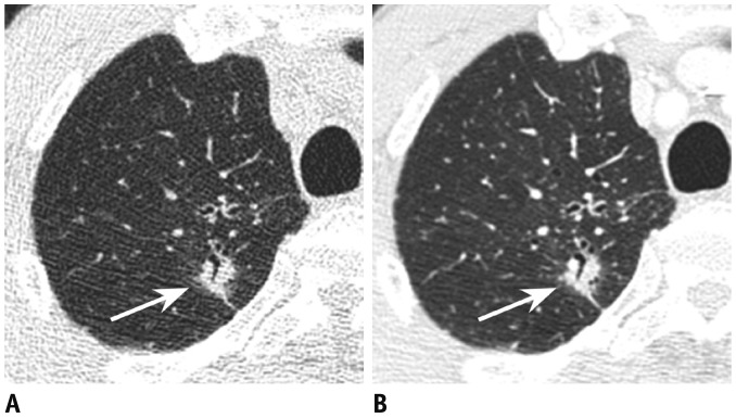
Materials and methods: From nodule libraries containing both low-dose unenhanced and standard-dose enhanced CT, 80 solid and 80 subsolid (40 part-solid, 40 non-solid) nodules of 135 patients were selected. Five thoracic radiologists categorized each nodule into solid, part-solid or non-solid. Inter-protocol agreement between low-dose unenhanced and standard-dose enhanced images was measured by pooling κ values for classification into two (solid, subsolid) and three (solid, part-solid, non-solid) categories. Interchangeability between low-dose unenhanced and standard-dose enhanced CT for the classification into two categories was assessed using a pre-defined equivalence limit of 8 percent.
Results: Inter-protocol agreement for the classification into two categories {κ, 0.96 (95% confidence interval [CI], 0.94-0.98)} and that into three categories (κ, 0.88 [95% CI, 0.85-0.92]) was considerably high. The probability of agreement between readers with standard-dose enhanced CT was 95.6% (95% CI, 94.5-96.6%), and that between low-dose unenhanced and standard-dose enhanced CT was 95.4% (95% CI, 94.7-96.0%). The difference between the two proportions was 0.25% (95% CI, -0.85-1.5%), wherein the upper bound CI was markedly below 8 percent.
Conclusion: Inter-protocol agreement for nodule classification was considerably high. Low-dose unenhanced CT can be used interchangeably with standard-dose enhanced CT for nodule classification.
Keywords: Classification; Computed tomography; Ground-glass nodule; Low-dose CT; Pulmonary nodules; Subsolid nodule.
Figures


Similar articles
-
Diameter of the Solid Component in Subsolid Nodules on Low-Dose Unenhanced Chest Computed Tomography: Measurement Accuracy for the Prediction of Invasive Component in Lung Adenocarcinoma.Korean J Radiol. 2018 May-Jun;19(3):508-515. doi: 10.3348/kjr.2018.19.3.508. Epub 2018 Apr 6. Korean J Radiol. 2018. PMID: 29713229 Free PMC article.
-
Observer Variability for Classification of Pulmonary Nodules on Low-Dose CT Images and Its Effect on Nodule Management.Radiology. 2015 Dec;277(3):863-71. doi: 10.1148/radiol.2015142700. Epub 2015 May 22. Radiology. 2015. PMID: 26020438
-
The effect of late-phase contrast enhancement on semi-automatic software measurements of CT attenuation and volume of part-solid nodules in lung adenocarcinomas.Eur J Radiol. 2016 Jun;85(6):1174-80. doi: 10.1016/j.ejrad.2016.03.027. Epub 2016 Mar 30. Eur J Radiol. 2016. PMID: 27161068
-
Overview and strategic management of subsolid pulmonary nodules.J Thorac Imaging. 2012 Jul;27(4):240-8. doi: 10.1097/RTI.0b013e31825d515b. J Thorac Imaging. 2012. PMID: 22847591 Review.
-
Update in the evaluation of the solitary pulmonary nodule.Radiographics. 2014 Oct;34(6):1658-79. doi: 10.1148/rg.346130092. Radiographics. 2014. PMID: 25310422 Review.
Cited by
-
A Glimpse on Trends and Characteristics of Recent Articles Published in the Korean Journal of Radiology.Korean J Radiol. 2019 Dec;20(12):1555-1561. doi: 10.3348/kjr.2019.0928. Korean J Radiol. 2019. PMID: 31854145 Free PMC article. No abstract available.
-
Improved repeatability of subsolid nodule measurement in low-dose lung screening with monoenergetic images: a phantom study.Quant Imaging Med Surg. 2019 Feb;9(2):171-179. doi: 10.21037/qims.2018.10.06. Quant Imaging Med Surg. 2019. PMID: 30976541 Free PMC article.
-
Retrospective Analysis of Subsolid Nodules' Frequency Using Chest Computed Tomography Detection in an Outpatient Population.Tomography. 2023 Aug 9;9(4):1494-1503. doi: 10.3390/tomography9040119. Tomography. 2023. PMID: 37624112 Free PMC article.
-
Comparative validation of clinical staging based on solid component versus total tumor size in resected lung adenocarcinoma.Eur Radiol. 2025 May 24. doi: 10.1007/s00330-025-11668-0. Online ahead of print. Eur Radiol. 2025. PMID: 40411551
References
-
- MacMahon H, Naidich DP, Goo JM, Lee KS, Leung AN, Mayo JR, et al. Guidelines for management of incidental pulmonary nodules detected on CT images: from the Fleischner Society 2017. Radiology. 2017;284:228–243. - PubMed
-
- American College of Radiology. Lung CT screening reporting and data system (Lung-RADS) [Accessed Apr 11, 2017]. Web site. https://www.acr.org/Clinical-Resources/Reporting-and-Data-Systems/Lung-Rads.
-
- Jacobs C, van Rikxoort EM, Scholten ET, de Jong PA, Prokop M, Schaefer-Prokop C, et al. Solid, part-solid, or non-solid?: classification of pulmonary nodules in low-dose chest computed tomography by a computer-aided diagnosis system. Invest Radiol. 2015;50:168–173. - PubMed
-
- van Riel SJ, Sanchez CI, Bankier AA, Naidich DP, Verschakelen J, Scholten ET, et al. Observer variability for classification of pulmonary nodules on low-dose CT images and its effect on nodule management. Radiology. 2015;277:863–871. - PubMed
Publication types
MeSH terms
LinkOut - more resources
Full Text Sources
Other Literature Sources
Medical
