

Quantitative proteomic analysis of pancreatic cyst fluid proteins associated with malignancy in intraductal papillary mucinous neoplasms
- PMID: 29713252
- PMCID: PMC5907296
- DOI: 10.1186/s12014-018-9193-1
Quantitative proteomic analysis of pancreatic cyst fluid proteins associated with malignancy in intraductal papillary mucinous neoplasms
Abstract
Background: The application of advanced imaging technologies for identifying pancreatic cysts has become widespread. However, accurately differentiating between low-grade dysplasia (LGD), high-grade dysplasia (HGD), and invasive intraductal papillary mucinous neoplasms (IPMNs) remains a diagnostic challenge with current biomarkers, necessitating the development of novel biomarkers that can distinguish IPMN malignancy.
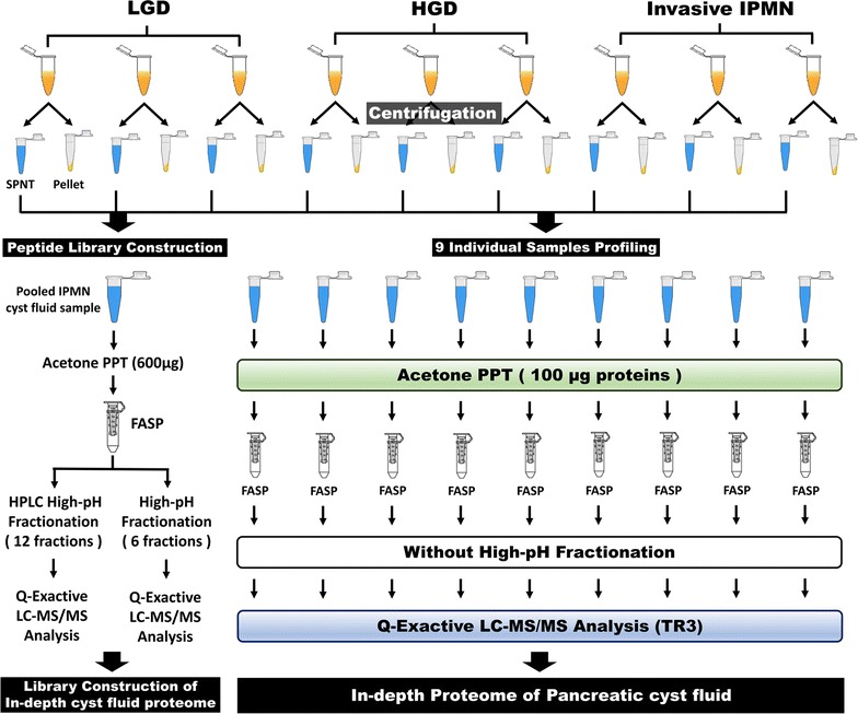
Methods: Cyst fluid samples were collected from nine IPMN patients (3 LGD, 3 HGD, and 3 invasive IPMN) during their pancreatectomies. An integrated proteomics approach that combines filter-aided sample preparation, stage tip-based high-pH fractionation, and high-resolution MS was applied to acquire in-depth proteomic data of pancreatic cyst fluid and discover marker candidates for IPMN malignancy. Biological processes of differentially expressed proteins that are related to pancreatic cysts and aggressive malignancy were analyzed using bioinformatics tools such as gene ontology analysis and Ingenuity pathway analysis. In order to confirm the validity of the marker candidates, 19 cyst fluid samples were analyzed by western blot.
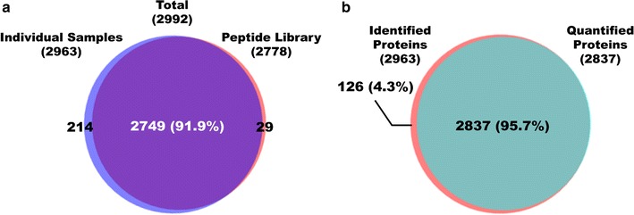
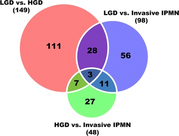
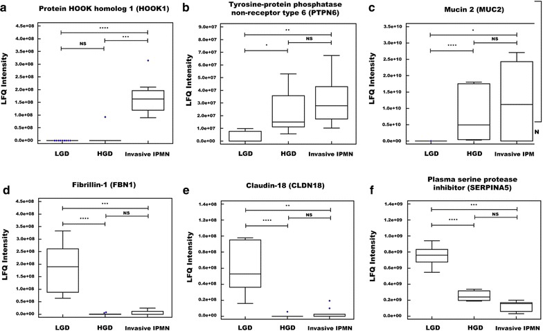
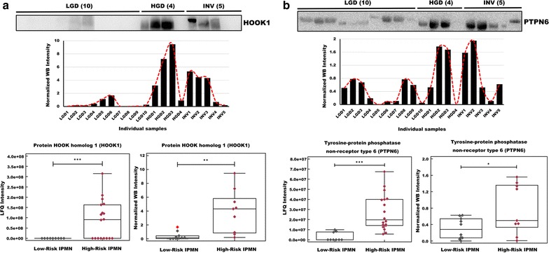
Results: A dataset of 2992 proteins was constructed from pancreatic cyst fluid samples. A subsequent analysis found 2963 identified proteins in individual samples, 2837 of which were quantifiable. Differentially expressed proteins between histological grades of IPMN were associated with pancreatic diseases and malignancy according to ingenuity pathway analysis. Eighteen biomarker candidates that were differentially expressed across IPMN histological grades were discovered-7 DEPs that were upregulated and 11 that were downregulated in more malignant grades. HOOK1 and PTPN6 were validated by western blot in an independent cohort, the results of which were consistent with our proteomic data.
Conclusions: This study demonstrates that novel biomarker candidates for IPMN malignancy can be discovered through proteomic analysis of pancreatic cyst fluid.
Keywords: Biomarker candidates; IPMN dysplasia; Intraductal papillary mucinous neoplasm (IPMN); LC–MS/MS; Pancreatic cyst fluid.
Figures


Similar articles
-
Marker Identification of the Grade of Dysplasia of Intraductal Papillary Mucinous Neoplasm in Pancreatic Cyst Fluid by Quantitative Proteomic Profiling.Cancers (Basel). 2020 Aug 23;12(9):2383. doi: 10.3390/cancers12092383. Cancers (Basel). 2020. PMID: 32842508 Free PMC article.
-
MUC2 expression and prevalence of high-grade dysplasia and invasive carcinoma in mixed-type intraductal papillary mucinous neoplasm of the pancreas.Pancreatology. 2013 Nov-Dec;13(6):583-8. doi: 10.1016/j.pan.2013.08.007. Epub 2013 Aug 30. Pancreatology. 2013. PMID: 24280573
-
Simultaneous and sequential combination of genetic and epigenetic biomarkers for the presence of high-grade dysplasia in patients with pancreatic cyst: Discovery in cyst fluid and test in pancreatic juice.Pancreatology. 2023 Mar;23(2):218-226. doi: 10.1016/j.pan.2023.01.006. Epub 2023 Jan 13. Pancreatology. 2023. PMID: 36707261
-
Humoral Predictors of Malignancy in IPMN: A Review of the Literature.Int J Mol Sci. 2021 Nov 27;22(23):12839. doi: 10.3390/ijms222312839. Int J Mol Sci. 2021. PMID: 34884643 Free PMC article. Review.
-
Cyst features and risk of malignancy in intraductal papillary mucinous neoplasms of the pancreas: a meta-analysis.Clin Gastroenterol Hepatol. 2013 Aug;11(8):913-21; quiz e59-60. doi: 10.1016/j.cgh.2013.02.010. Epub 2013 Feb 13. Clin Gastroenterol Hepatol. 2013. PMID: 23416279 Review.
Cited by
-
Translation of a Protease Turnover Assay for Clinical Discrimination of Mucinous Pancreatic Cysts.Diagnostics (Basel). 2022 May 28;12(6):1343. doi: 10.3390/diagnostics12061343. Diagnostics (Basel). 2022. PMID: 35741154 Free PMC article.
-
Proteogenomic analysis reveals Arp 2/3 complex as a common molecular mechanism in high risk pancreatic cysts and pancreatic cancer.Sci Rep. 2025 Jan 31;15(1):3902. doi: 10.1038/s41598-025-87872-1. Sci Rep. 2025. PMID: 39890846 Free PMC article.
-
Molecular Diagnosis of Cystic Neoplasms of the Pancreas: a Review.J Gastrointest Surg. 2020 May;24(5):1201-1214. doi: 10.1007/s11605-020-04537-2. Epub 2020 Mar 3. J Gastrointest Surg. 2020. PMID: 32128679 Review.
-
Thermal Liquid Biopsy (TLB) Focused on Benign and Premalignant Pancreatic Cyst Diagnosis.J Pers Med. 2020 Dec 31;11(1):25. doi: 10.3390/jpm11010025. J Pers Med. 2020. PMID: 33396529 Free PMC article.
-
Marker Identification of the Grade of Dysplasia of Intraductal Papillary Mucinous Neoplasm in Pancreatic Cyst Fluid by Quantitative Proteomic Profiling.Cancers (Basel). 2020 Aug 23;12(9):2383. doi: 10.3390/cancers12092383. Cancers (Basel). 2020. PMID: 32842508 Free PMC article.
References
-
- Larghi A, Panic N, Capurso G, Leoncini E, Arzani D, Salvia R, Del Chiaro M, Frulloni L, Arcidiacono PG, Zerbi A, et al. Prevalence and risk factors of extrapancreatic malignancies in a large cohort of patients with intraductal papillary mucinous neoplasm (IPMN) of the pancreas. Ann Oncol. 2013;24:1907–1911. - PubMed
-
- Hwang DW, Jang JY, Lee SE, Lim CS, Lee KU, Kim SW. Clinicopathologic analysis of surgically proven intraductal papillary mucinous neoplasms of the pancreas in SNUH: a 15-year experience at a single academic institution. Langenbecks Arch Surg. 2012;397:93–102. - PubMed
-
- Gourgiotis S, Ridolfini MP, Germanos S. Intraductal papillary mucinous neoplasms of the pancreas. Eur J Surg Oncol. 2007;33:678–684. - PubMed
-
- Tanaka M, Fernandez-del Castillo C, Adsay V, Chari S, Falconi M, Jang JY, Kimura W, Levy P, Pitman MB, Schmidt CM, et al. International consensus guidelines 2012 for the management of IPMN and MCN of the pancreas. Pancreatology. 2012;12:183–197. - PubMed
LinkOut - more resources
Full Text Sources
Other Literature Sources