A Case of Nongerminomatous Germ Cell Tumor of the Pineal Region: Risks and Advantages of Biopsy by Endoscopic Approach
- PMID: 29713348
- PMCID: PMC5866897
- DOI: 10.1155/2018/5106701
A Case of Nongerminomatous Germ Cell Tumor of the Pineal Region: Risks and Advantages of Biopsy by Endoscopic Approach
Abstract
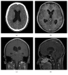
A 21-year-old male was admitted to our department with headache and drowsiness. CT scan and MRI revealed acute obstructive hydrocephalus caused by a pineal region mass. The serum and CSF levels of beta-human chorionic gonadotropin (beta-hCG) were 215 IU/L and 447 IU/L, respectively, while levels of alpha-fetoprotein (AFP) were normal. A germ cell tumor (GCT) was suspected, and the patient underwent endoscopic third ventriculostomy (ETV) with biopsy. After four days from surgery, the tumor bled with mass expansion and ETV stoma occlusion; thus, a ventriculoperitoneal shunt was positioned. After ten months, the tumor metastasized to the thorax and abdomen with progression of intracerebral tumor mass. Despite the aggressive nature of this tumor, ETV remains a valid approach for a pineal region mass, but in case of GCT, the risk of bleeding should be taken into account, during and after the surgical procedure.
Figures


Similar articles
-
Mixed germ cell tumor infiltrating the pineal gland without elevated tumor markers: illustrative case.J Neurosurg Case Lessons. 2021 Mar 22;1(12):CASE20131. doi: 10.3171/CASE20131. eCollection 2021 Mar 22. J Neurosurg Case Lessons. 2021. PMID: 35854926 Free PMC article.
-
[Endoscopic treatment strategy of obstructive hydrocephalus induced by pineal region tumors].Zhonghua Yi Xue Za Zhi. 2015 Mar 24;95(11):845-8. Zhonghua Yi Xue Za Zhi. 2015. PMID: 26080918 Chinese.
-
Outcomes in patients undergoing single-trajectory endoscopic third ventriculostomy and endoscopic biopsy for midline tumors presenting with obstructive hydrocephalus.J Neurosurg. 2006 Sep;105(3 Suppl):219-26. doi: 10.3171/ped.2006.105.3.219. J Neurosurg. 2006. PMID: 16970236
-
Pineal region tumors: simultaneous endoscopic third ventriculostomy and tumor biopsy.World Neurosurg. 2013 Feb;79(2 Suppl):S18.e9-13. doi: 10.1016/j.wneu.2012.02.020. Epub 2012 Feb 10. World Neurosurg. 2013. PMID: 22381843 Review.
-
Outcome of single-trajectory rigid endoscopic third ventriculostomy and biopsy in the management algorithm of pineal region tumors: a case series and review of the literature.Childs Nerv Syst. 2018 Jul;34(7):1335-1344. doi: 10.1007/s00381-018-3840-8. Epub 2018 May 28. Childs Nerv Syst. 2018. PMID: 29808320 Review.
Cited by
-
Surgical strategy for symptomatic pineal cyst: is endoscopit third ventriculostomy necessary in addition to cyst fenestration?Nagoya J Med Sci. 2021 Aug;83(3):627-633. doi: 10.18999/nagjms.83.3.627. Nagoya J Med Sci. 2021. PMID: 34552294 Free PMC article.
References
-
- The Committee of Brain Tumor Registry of Japan. Special report of brain tumor registry of Japan (1969–1993) Neurologia Medico-Chirurgica. 2000;40(9-10) - PubMed
Publication types
LinkOut - more resources
Full Text Sources
Other Literature Sources

