Intestinal duplication revealed by posterior reversible encephalopathy syndrome
- PMID: 29713360
- PMCID: PMC5924845
- DOI: 10.3345/kjp.2018.61.4.132
Intestinal duplication revealed by posterior reversible encephalopathy syndrome
Abstract
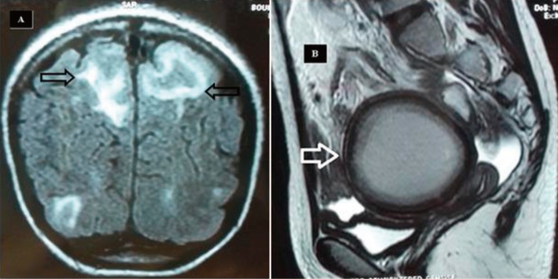
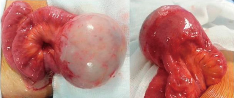
We report a unique case of intestinal duplication detected on posterior reversible encephalopathy syndrome (PRES) in a 13-year-old girl. She was admitted to the pediatric Emergency Department because of generalized seizures. Radiological assessment revealed a large, well-defined, thick-walled cystic lesion in the mid abdomen, suggestive of duplication cyst associated to a PRES. Exploration confirmed the diagnosis of ileal duplication cyst, and the mass was resected. The postoperative course was uneventful. Both hypertension and neurological dysfunction resolved after the mass resection. A followup brain magnetic resonance imaging was performed 9 months later and showed complete resolution of the cerebellar changes. Although extrinsic compression of the retroperitoneal structures has not been reported in the literature as a complication of duplication cyst, we strongly believe that this is the most logical and plausible hypothesis that would explain the pathogenesis of PRES in our patient.
Keywords: Cyst; Hypertension; Ileum; Metoclopramide; Posterior Reversible Encephalopathy Syndrome.
Conflict of interest statement
Conflicts of interest: No potential conflict of interest relevant to this article was reported.
Figures
Similar articles
-
Posterior reversible encephalopathy syndrome (PRES): a rare condition after resection of posterior fossa tumors: two new cases and review of the literature.Childs Nerv Syst. 2016 May;32(5):857-63. doi: 10.1007/s00381-015-2954-5. Epub 2015 Nov 19. Childs Nerv Syst. 2016. PMID: 26584552 Review.
-
Sequential occurrence of eclampsia-associated posterior reversible encephalopathy syndrome and reversible splenial lesion syndrome (a case report): proposal of a novel pathogenesis for reversible splenial lesion syndrome.BMC Med Imaging. 2019 Apr 30;19(1):35. doi: 10.1186/s12880-019-0323-7. BMC Med Imaging. 2019. PMID: 31039748 Free PMC article.
-
Posterior reversible encephalopathy syndrome (PRES) induced by pazopanib, a multi-targeting tyrosine kinase inhibitor, in a patient with soft-tissue sarcoma: case report and review of the literature.Invest New Drugs. 2018 Apr;36(2):346-349. doi: 10.1007/s10637-017-0521-5. Epub 2017 Oct 25. Invest New Drugs. 2018. PMID: 29067537 Free PMC article. Review.
-
Posterior reversible encephalopathy syndrome following elevated mean arterial pressures for cervical spinal cord injury.J Spinal Cord Med. 2018 Jan;41(1):111-114. doi: 10.1080/10790268.2016.1250030. Epub 2016 Dec 5. J Spinal Cord Med. 2018. PMID: 27917700 Free PMC article.
-
Reversible posterior encephalopathy syndrome in a 10-year-old child.J Bras Nefrol. 2019 Jul-Sep;41(3):436-439. doi: 10.1590/2175-8239-JBN-2018-0111. Epub 2018 Sep 21. J Bras Nefrol. 2019. PMID: 30281063 Free PMC article.
Cited by
-
Can Post-Operative Posterior Reversible Encephalopathy Syndrome (PRES) Be Considered an Insidious Rare Surgical Complication?Brain Sci. 2023 Apr 23;13(5):706. doi: 10.3390/brainsci13050706. Brain Sci. 2023. PMID: 37239179 Free PMC article. Review.
References
-
- Gümüş H, Per H, Kumandaş S, Yikilmaz A. Reversible posterior leukoencephalopathy syndrome in childhood: report of nine cases and review of the literature. Neurol Sci. 2010;31:125–131. - PubMed
-
- Zuccoli G, Fitzgerald RT, Nardone R, Furtado AD, Abdel-Hamid H. The link between arterial blood pressure and vasogenic edema in pediatric PRES. Neuroradiology. 2015;57:865–866. - PubMed
LinkOut - more resources
Full Text Sources
Other Literature Sources