. 2017 Oct;1(2):231-241.
doi: 10.1002/rth2.12034. Epub 2017 Aug 10.

Clot stability as a determinant of effective factor VIII replacement in hemophilia A

Affiliations

Clot stability as a determinant of effective factor VIII replacement in hemophilia A

L Leong et al. Res Pract Thromb Haemost. 2017 Oct.

Abstract

Background: Factor VIII (FVIII) replacement is standard of care for patients with hemophilia A (HemA); however, patient response does not always correlate with FVIII levels. We hypothesize this may be in part due to the physical properties of clots and contributions of fibrin, platelets, and erythrocytes, which may be important for hemostasis.

Objective: To understand how FVIII contributes to effective hemostasis in terms of clot structure and mechanical properties.

Patients/methods: In vitro HemA clots in human plasma or whole blood were analyzed using turbidity waveform analysis, confocal microscopy, and rheometry with or without added FVIII. In vivo clots from saphenous vein puncture in wild-type and HemA mice with varying FVIII levels were examined using scanning electron microscopy.

Results: FVIII profoundly affected HemA clot structure and physical properties; added FVIII converted the open and porous fibrin meshwork and low stiffness of HemA clots to a highly branched and dense meshwork with higher stiffness. Platelets and erythrocytes incorporated into clots modulated clot properties. The clots formed in the mouse saphenous vein model contained variable amounts of compressed erythrocytes (polyhedrocytes), fibrin, and platelets depending on the levels of FVIII, correlating with bleeding times. FVIII effects on clot characteristics were dose-dependent and reached a maximum at ~25% FVIII, such that HemA clots formed with this level of FVIII resembled clots from unaffected controls.

Conclusions: Effective clot formation can be achieved in HemA by replacement therapy, which alters the architecture of the fibrin network and associated cells, thus increasing clot stiffness and decreasing clot permeability.

Keywords: blood coagulation; factor VIII; fibrin; hemophilia A; thrombin.


Conflict of interest statement

Conflict-of-interest disclosure: I. N. Chernysh, C. Nagaswami, Z. de Lange, S. Kosolapova, and A. Cuker have no conflicts of interest to disclose.

Figures

Figure 1
Figure 1
Turbidity profiles of HemA plasma clots with and without FVIII supplementation. The heterogeneity of individual HemA plasma is evident in the (A) CTs, (B) fibrinogen levels, and (C) maximum turbidity, which is directly proportional to the fibrin fiber cross‐sectional area.17 The correlation between fibrinogen and maximum turbidity is evident in the r 2 of ~1. Fibrinogen levels of normal pool in panel B are indicated with a green arrow. (D) Clot time and turbidity maxima of HemA donors with FVIII supplementation. Individual HemA plasma samples are denoted by different symbols: without FVIII (red outline) or with spikes of 1% FVIII (orange outline), 5% FVIII (blue outline), or 25% FVIII (cyan outline). aPTT, activated partial thromboplastin time; CT, clot time; FVIII, factor VIII; HemA, hemophilia A
Figure 2
Figure 2
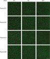
Effect of FVIII on HemA plasma clot structure. Changes in individual HemA plasma clot structure with the indicated FVIII supplementation were visualized by confocal microscopy as described in the Methods. Levels of FVIII depicted were chosen to capture potential clot structures formed in severe (0%) and mild (5% and 25%) HemA. Effects of other FVIII levels reflective of the continuum of HemA disease severity can be further visualized in Fig. S2. rFVIII, recombinant FVIII
Figure 3
Figure 3
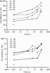
FVIII‐mediated changes in whole blood rheological properties in HemA. (A and B) Rheology results of whole blood clots derived from individual HemA blood are indicated by different open symbols. Note that although FVIII increased both clot stiffness and clot plasticity, the individual HemA blood samples retained, in general, their baseline (no FVIII) rank orders, suggesting maintenance of individual characteristics. FVIII, factor VIII; HemA, hemophilia A
Figure 4
Figure 4
Saphenous vein injury model in HemA mice. HemA mice were given rFVIII such that at the time of vascular injury, the plasma FVIII level was predicted by pharmacokinetics to be at the desired level. (A) Vascular injury induction. (B) Bleeding times recorded for HemA mice dosed with the different FVIII (n=5/dose). Note that the bleeding time of HemA mice with 25% and 100% plasma FVIII were essentially comparable when the variance in bleeding time was taken into account. In these studies, C57BL mice treated with excipient (n=5) served as normal controls. FVIII, factor VIII; HemA, hemophilia A
Figure 5
Figure 5
Structure of typical mouse saphenous vein clots. Scanning electron micrographs of the most typical areas of wild‐type mouse saphenous vein clots. (A) Closely packed polyhedrocytes surrounded by a meshwork of fibrin and platelets. The top surface is mostly polyhedrocytes, and the sides are a dense mesh of fibrin and platelets. (B) Higher magnification view of the upper left part of the top surface, showing the tightly packed polyhedrocytes. (C) Higher magnification view of lower central part of the top surface, showing more polyhedrocytes. (D) Higher magnification view of another clot with closely packed polyhedrocytes and fibrin and platelets on the left. (E) Another clot with polyhedrocytes on the right and fibrin mesh with platelets on the left, including tethered balloon‐like erythrocytes. (F) Another clot with fibrin mesh with platelets on the top, including tethered balloon‐like erythrocytes, and polyhedrocytes below. Magnification bars for all panels=10 μm
Figure 6
Figure 6
Structures identified in hemophilia A (HemA) saphenous vein clots. (A–C [16 images with 9796 structures]) Clots from HemA mice with low levels of plasma FVIII. (D–F) Clots from HemA mice with higher levels of FVIII (25%: D–E [11 images with 13,894 structures]; 100%: F [17 images with 14 059 structures]). (G–I) Clots derived from HemA mice with (G) 100% FVIII and from (H and I [16 images with 18 227 structures]) control, wild‐type mice. (A) HemA mouse with 5% FVIII. Mostly platelets, some fibrin. (B) HemA mouse with 5% FVIII. Dense contracted clot with platelets and fibrin on the outside. (C) HemA mouse with 5% FVIII. Polyhedrocytes and fibrin. (D) HemA mouse with 25% FVIII. Fibrin and platelets with some intermediate forms of polyhedrocytes. (E) HemA mouse with 25% FVIII. Mostly aggregated platelets. (F) HemA mouse with 100% FVIII. Fibrin and platelets. (G) HemA mouse with 100% FVIII. Red blood cell balloons trapped in fibrin mesh. (H) Control mouse. Polyhedrocytes and fibrin mesh. (I) Control mouse. Mostly fibrin with a few platelets. Magnification bar for all panels=10 μm
Figure 7
Figure 7
Quantitation of structures identified in saphenous vein clots. Saphenous vein clots visualized using scanning electron micrographs were quantified as described in the Section 2. The proportion of total visual field occupied by these structures were averaged. The fibrin and erythrocyte (polyhedrocyte as well as combined intermediate form and polyhedrocyte) structures identified in clots derived from HemA mice containing 5%, 25%, and 100% FVIII or wild‐type C57BL mice are normalized against the platelet. Quantitative changes induced by FVIII were compared statistically by ANOVA test with Dunnett correction for multiple comparisons, and the levels of statistical significance for each comparison were done with a multiple Student t test. P values for differences between 5% FVIII, 25% FVIII, 100% FVII, and control are indicated. (A) Fibrin component. (B) Polyhedral‐shaped erythrocytes. (C) Combination of polyhedrocytes and intermediate erythrocytes. ANOVA, analysis of variance; FVIII, factor VII

Similar articles

Cited by

References

    1. Srivastava A, Brewer AK, Mauser‐Bunschoten EP, et al. ; Treatment Guidelines Working Group on behalf of the World Federation Of Hemophilia . Guidelines for the management of hemophilia. Haemophilia. 2013;19:e1–47. - PubMed
    1. Hilden I, Lauritzen B, Sorensen BB, et al. Hemostatic effect of a monoclonal antibody mAb 2021 blocking the interaction between FXa and TFPI in a rabbit hemophilia model. Blood. 2012;119:5871–8. - PubMed
    1. Sehgal A, Barros S, Ivanciu L, et al. An RNAi therapeutic targeting antithrombin to rebalance the coagulation system and promote hemostasis in hemophilia. Nat Med. 2015;21:492–7. - PubMed
    1. Muto A, Yoshihashi K, Takeda M, et al. Anti‐factor IXa/X bispecific antibody (ACE910): hemostatic potency against ongoing bleeds in a hemophilia A model and the possibility of routine supplementation. J Thromb Haemost. 2014;12:206–13. - PubMed
    1. Srivastava A. Haemophilia care ‐ beyond the treatment guidelines. Haemophilia. 2014;20(Suppl 4):4–10. - PubMed