Multicenter Study
. 2018 Jul;288(1):26-35.
doi: 10.1148/radiol.2018172462. Epub 2018 May 1.

Intratumoral Spatial Heterogeneity at Perfusion MR Imaging Predicts Recurrence-free Survival in Locally Advanced Breast Cancer Treated with Neoadjuvant Chemotherapy

Affiliations
Multicenter Study

Intratumoral Spatial Heterogeneity at Perfusion MR Imaging Predicts Recurrence-free Survival in Locally Advanced Breast Cancer Treated with Neoadjuvant Chemotherapy

Jia Wu et al. Radiology. 2018 Jul.

Abstract

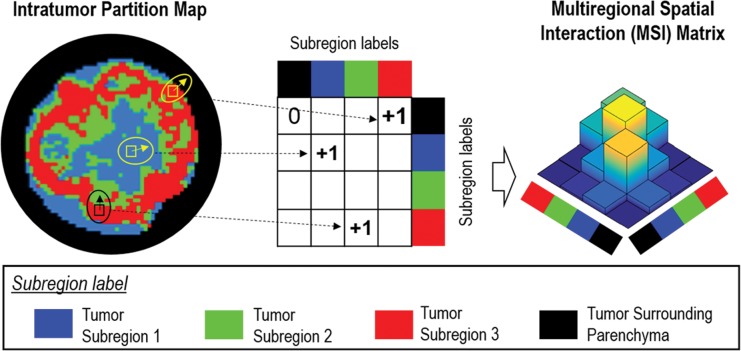
Purpose To characterize intratumoral spatial heterogeneity at perfusion magnetic resonance (MR) imaging and investigate intratumoral heterogeneity as a predictor of recurrence-free survival (RFS) in breast cancer. Materials and Methods In this retrospective study, a discovery cohort (n = 60) and a multicenter validation cohort (n = 186) were analyzed. Each tumor was divided into multiple spatially segregated, phenotypically consistent subregions on the basis of perfusion MR imaging parameters. The authors first defined a multiregional spatial interaction (MSI) matrix and then, based on this matrix, calculated 22 image features. A network strategy was used to integrate all image features and classify patients into different risk groups. The prognostic value of imaging-based stratification was evaluated in relation to clinical-pathologic factors with multivariable Cox regression. Results Three intratumoral subregions with high, intermediate, and low MR perfusion were identified and showed high consistency between the two cohorts. Patients in both cohorts were stratified according to network analysis of multiregional image features regarding RFS (log-rank test, P = .002 for both). Aggressive tumors were associated with a larger volume of the poorly perfused subregion as well as interaction between poorly and moderately perfused subregions and surrounding parenchyma. At multivariable analysis, the proposed MSI-based marker was independently associated with RFS (hazard ratio: 3.42; 95% confidence interval: 1.55, 7.57; P = .002) adjusting for age, estrogen receptor (ER) status, progesterone receptor status, human epidermal growth factor receptor type 2 (HER2) status, tumor volume, and pathologic complete response (pCR). Furthermore, imaging helped stratify patients for RFS within the ER-positive and HER2-positive subgroups (log-rank test, P = .007 and .004) and among patients without pCR after neoadjuvant chemotherapy (log-rank test, P = .003). Conclusion Breast cancer consists of multiple spatially distinct subregions. Imaging heterogeneity is an independent prognostic factor beyond traditional risk predictors.


Figures

Figure 1:
Figure 1:
Flowchart shows number of patients with breast cancer in two study cohorts. DCE = dynamic contrast-enhanced, I-SPY 1 TRIAL = Investigation of Serial Studies to Predict Your Therapeutic Response with Imaging and Molecular Analysis, UCSF = University of California, San Francisco.
Figure 2a:
Figure 2a:
(a) Proposed two-stage intratumor partition framework. DCE = dynamic contrast-enhanced. (b) Illustration shows use of multiregional spatial interaction (MSI) matrix derived from intratumor partition maps. (c) Twenty-two quantitative imaging features were extracted from MSI matrix to measure intratumoral spatial heterogeneity.
Figure 2b:
Figure 2b:
(a) Proposed two-stage intratumor partition framework. DCE = dynamic contrast-enhanced. (b) Illustration shows use of multiregional spatial interaction (MSI) matrix derived from intratumor partition maps. (c) Twenty-two quantitative imaging features were extracted from MSI matrix to measure intratumoral spatial heterogeneity.
Figure 2c:
Figure 2c:
(a) Proposed two-stage intratumor partition framework. DCE = dynamic contrast-enhanced. (b) Illustration shows use of multiregional spatial interaction (MSI) matrix derived from intratumor partition maps. (c) Twenty-two quantitative imaging features were extracted from MSI matrix to measure intratumoral spatial heterogeneity.
Figure 3:
Figure 3:
Box-and-whisker plots show distribution of four perfusion imaging parameters for three intratumoral subregions for, A–D, cohort 1 and, E–H, cohort 2. PE = percentage enhancement, SER = signal enhancement ratio, WIS = wash-in slope, WOS = washout slope. P values were obtained with Student t test. * = P < .05, ** = P < .001, *** = P < .0001.
Figure 4:
Figure 4:
A, Sparse graph with seven neighbors shows 60 patients in discovery cohort after proposed network analysis. Two clusters were identified, with 21 patients in one cluster labeled in red and remaining 39 patients in another cluster labeled in green. Each vertex represents an individual patient, and its size is proportional to tumor volume. B, Kaplan-Meier curves of recurrence-free survival stratified according to the two patient clusters. Coloring is consistent with patient cluster in A.
Figure 5:
Figure 5:
Kaplan-Meier curves of recurrence-free survival in validation cohort. Patients are stratified according to propagated patient cluster labels in discovery cohort (Fig 3, A). Plots are for, A, entire validation cohort (Investigation of Serial Studies to Predict Your Therapeutic Response with Imaging and Molecular Analysis), B, estrogen receptor–positive subgroup, C, human epidermal growth factor receptor type 2–positive subgroup, and, D, subgroup showing no pathologic complete response. HR = hazard ratio.
Figure 6:
Figure 6:
Intratumor partition maps in two breast cancer patients. The proposed analysis pipeline accurately predicted their recurrence risk. High-perfusion subregion is in red, with intermediate perfusion in green and low perfusion in blue. ER = estrogen receptor, HER2 = human epidermal growth factor receptor type 2, LN = lymph node, MSI = multiregional spatial interaction.

Comment in

References

    1. Gralow JR, Burstein HJ, Wood W, et al. . Preoperative therapy in invasive breast cancer: pathologic assessment and systemic therapy issues in operable disease. J Clin Oncol 2008;26(5):814–819. - PubMed
    1. Esserman LJ, Berry DA, DeMichele A, et al. . Pathologic complete response predicts recurrence-free survival more effectively by cancer subset: results from the I-SPY 1 TRIAL—CALGB 150007/150012, ACRIN 6657. J Clin Oncol 2012;30(26):3242–3249. - PMC - PubMed
    1. Hylton NM, Gatsonis CA, Rosen MA, et al. . Neoadjuvant chemotherapy for breast cancer: functional tumor volume by MR imaging predicts recurrence-free survival—results from the ACRIN 6657/CALGB 150007 I-SPY 1 TRIAL. Radiology 2016;279(1):44–55. - PMC - PubMed
    1. Esserman LJ, Thompson IM, Reid B, et al. . Addressing overdiagnosis and overtreatment in cancer: a prescription for change. Lancet Oncol 2014;15(6):e234–e242. - PMC - PubMed
    1. Zardavas D, Irrthum A, Swanton C, Piccart M. Clinical management of breast cancer heterogeneity. Nat Rev Clin Oncol 2015;12(7):381–394. - PubMed

Publication types