Review
. 2018 May 1;128(5):1711-1719.
doi: 10.1172/JCI120330. Epub 2018 May 1.

Interpreting heterogeneity in intestinal tuft cell structure and function

Affiliations
Review

Interpreting heterogeneity in intestinal tuft cell structure and function

Amrita Banerjee et al. J Clin Invest. .

Abstract

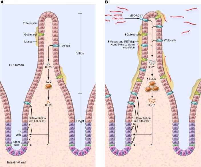
Intestinal tuft cells are a morphologically unique cell type, best characterized by striking microvilli that form an apical tuft. These cells represent approximately 0.5% of gut epithelial cells depending on location. While they are known to express chemosensory receptors, their function has remained unclear. Recently, numerous groups have revealed startling insights into intestinal tuft cell biology. Here, we review the latest developments in understanding this peculiar cell type's structure and function. Recent advances in volumetric microscopy have begun to elucidate tuft cell ultrastructure with respect to its cellular neighbors. Moreover, single-cell approaches have revealed greater diversity in the tuft cell population than previously appreciated and uncovered novel markers to characterize this heterogeneity. Finally, advanced model systems have revealed tuft cells' roles in mucosal healing and orchestrating type 2 immunity against eukaryotic infection. While much remains unknown about intestinal tuft cells, these critical advances have illuminated the physiological importance of these previously understudied cells and provided experimentally tractable tools to interrogate this rare cell population. Tuft cells act as luminal sensors, linking the luminal microbiome to the host immune system, which may make them a potent clinical target for modulating host response to a variety of acute or chronic immune-driven conditions.


Conflict of interest statement

Conflict of interest: The authors have declared that no conflict of interest exists.

Figures

Figure 1
Figure 1. Structural characteristics of intestinal tuft or caveolated cells.
Intestinal tuft cells are easily distinguishable from neighboring enterocytes by their unique apical brush border and oval-shaped cell body. The apical microvilli connect the extracellular environment of the lumen to the intracellular cytoplasm via a filamentous bundle. Vesicles carrying unknown cargo are interspersed within the filamentous bundle, which terminates at a tubular network at the apex of the tuft cell nucleus. Lateral membrane projections or cytospinules emanate from the tuft cell and pierce the membrane of adjacent enterocytes. Cytospinules have been shown to directly contact the nuclei of tuft cell neighbors, possibly serving as a means of cell-to-cell communication.
Figure 2
Figure 2. Small intestinal tuft cells induce a type 2 immune response following eukaryotic colonization of the gut.
At homeostasis, DCLK1+ cells promote their own specification through release of IL-25, which stimulates ILC2s to produce IL-13. By an unknown mechanism, IL-13 stimulates stem cells and/or transit-amplifying progenitors to differentiate into tuft cells. During eukaryotic colonization of the small intestine, tuft cells sense pathogens through an ill-defined mechanism, possibly involving the mTORC1 complex and Raptor. Increased IL-25 release by tuft cells drives IL-13–dependent expansion of the tuft cell lineage. Goblet cell hyperplasia and increased release of mucus as well as the anti-helminth molecule RETN1β contribute to worm expulsion from the proximal gut.

References

    1. Luciano L, Reale E. A new morphological aspect of the brush cells of the mouse gallbladder epithelium. Cell Tissue Res. 1979;201(1):37–44. - PubMed
    1. Luciano L, Reale E. Brush cells of the mouse gallbladder. A correlative light- and electron-microscopical study. Cell Tissue Res. 1990;262(2):339–349. doi: 10.1007/BF00309889. - DOI - PubMed
    1. Luciano L, Reale E. Presence of brush cells in the mouse gallbladder. Microsc Res Tech. 1997;38(6):598–608. doi: 10.1002/(SICI)1097-0029(19970915)38:6<598::AID-JEMT4>3.0.CO;2-B. - DOI - PubMed
    1. Luciano L, Groos S, Reale E. Brush cells of rodent gallbladder and stomach epithelia express neurofilaments. J Histochem Cytochem. 2003;51(2):187–198. doi: 10.1177/002215540305100207. - DOI - PubMed
    1. Gilloteaux J, Pomerants B, Kelly TR. Human gallbladder mucosa ultrastructure: evidence of intraepithelial nerve structures. Am J Anat. 1989;184(4):321–333. doi: 10.1002/aja.1001840407. - DOI - PubMed

Publication types