. 2018 May 1;6(1):35.
doi: 10.1186/s40478-018-0537-x.

α-Synuclein fibril-induced paradoxical structural and functional defects in hippocampal neurons

Affiliations

α-Synuclein fibril-induced paradoxical structural and functional defects in hippocampal neurons

Jessica M Froula et al. Acta Neuropathol Commun. .

Abstract

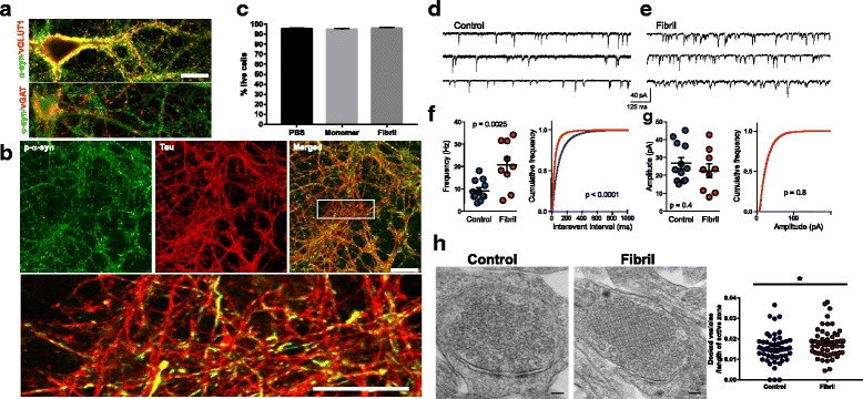
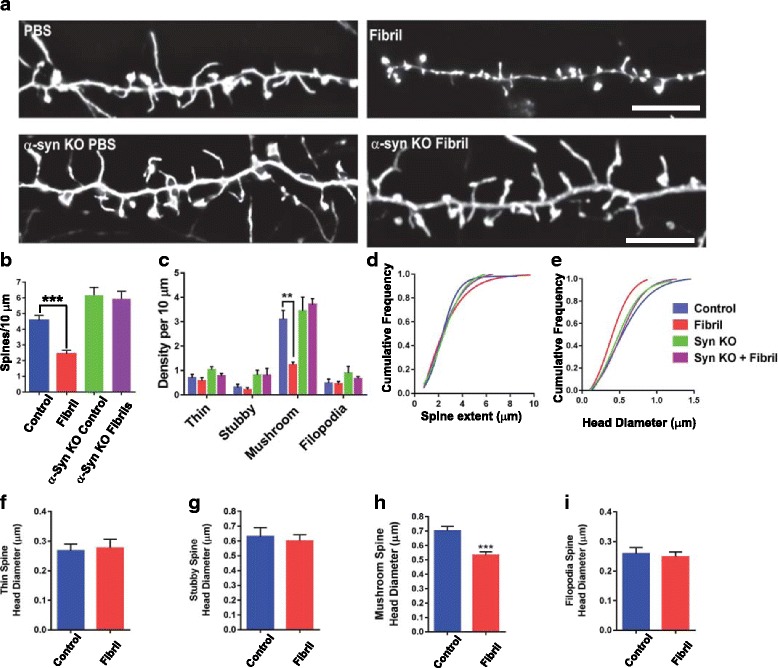
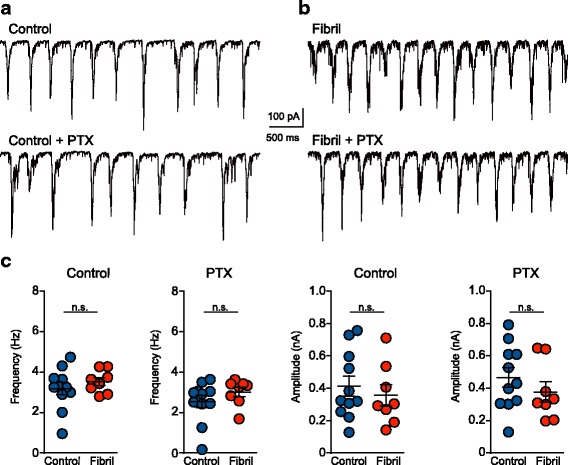
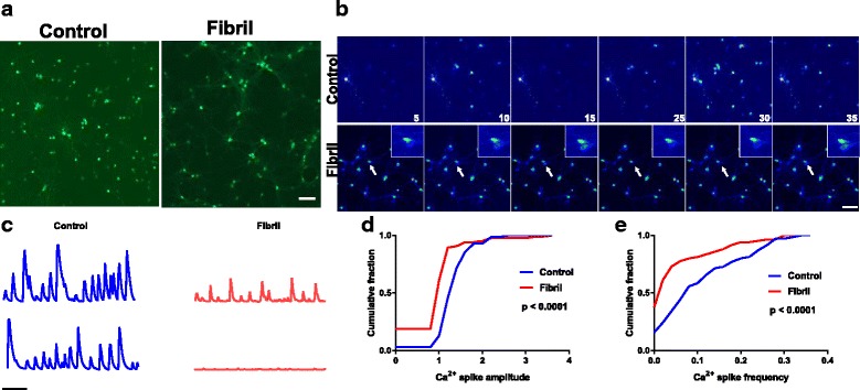
Neuronal inclusions composed of α-synuclein (α-syn) characterize Parkinson's Disease (PD) and Dementia with Lewy bodies (DLB). Cognitive dysfunction defines DLB, and up to 80% of PD patients develop dementia. α-Syn inclusions are abundant in the hippocampus, yet functional consequences are unclear. To determine if pathologic α-syn causes neuronal defects, we induced endogenous α-syn to form inclusions resembling those found in diseased brains by treating hippocampal neurons with α-syn fibrils. At seven days after adding fibrils, α-syn inclusions are abundant in axons, but there is no cell death at this time point, allowing us to assess for potential alterations in neuronal function that are not caused by neuron death. We found that exposure of neurons to fibrils caused a significant reduction in mushroom spine densities, adding to the growing body of literature showing that altered spine morphology is a major pathologic phenotype in synucleinopathies. The reduction in spine densities occurred only in wild type neurons and not in neurons from α-syn knockout mice, suggesting that the changes in spine morphology result from fibril-induced corruption of endogenously expressed α-syn. Paradoxically, reduced postsynaptic spine density was accompanied by increased frequency of miniature excitatory postsynaptic currents (EPSCs) and presynaptic docked vesicles, suggesting enhanced presynaptic function. Action-potential dependent activity was unchanged, suggesting compensatory mechanisms responding to synaptic defects. Although activity at the level of the synapse was unchanged, neurons exposed to α-syn fibrils, showed reduced frequency and amplitudes of spontaneous Ca2+ transients. These findings open areas of research to determine the mechanisms that alter neuronal function in brain regions critical for cognition at time points before neuron death.

Keywords: Calcium imaging; Dendritic spines; Fibril; Lewy body; Lewy neurite; Parkinson’s disease; Physiology; α-Synuclein.


Conflict of interest statement

Ethics approval and consent to participate

Not applicable.

Competing interests

The authors declare that they have no competing interests.

Publisher’s Note

Springer Nature remains neutral with regard to jurisdictional claims in published maps and institutional affiliations.

Figures

Fig. 1
Fig. 1
Seven days after exposure to α-syn fibrils, α-syn inclusions localize to axons, increase mEPSC frequency and increase the number of docked presynaptic vesicles. a Untreated primary hippocampal neurons were fixed at DIV 14. Immunofluorescence was performed with antibodies to total α-syn (green) and vGLuT1 (red) to label glutamatergic presynaptic terminals or vGAT1 (red) to label GABAergic terminals. Scale bar = 10 μm. b, c α-syn fibrils (2 μg/mL) were added to primary hippocampal neurons on DIV 7 and neurons were fixed 7 days later (DIV14). Immunofluorescence was performed using antibodies to p-α-syn (green) to label inclusions or tau (red) to label axons. Images were captured with a confocal microscope at an optical thickness of 0.5 μm. Scale bar = 50 μm, top panel, 25 μm, bottom panel. c Primary neurons were exposed to 2 μg/mL monomer, 2 μg/mL fibrils or PBS at DIV 7. Seven days later (DIV14), calcein AM was used to label live cells and ethidium homodimer-1 was used to label dead cells. Each well was scanned and tiled at 10X. Image J was used to quantify live and dead cells. A total of 32,527 PBS treated cells, 40,248 monomer treated cells, and 39,105 fibril treated cells were counted in two independent experiments. Data is expressed as the average live cells/total number of cells (sum of calcein positive and ethidium homodimer positive). p = 0.864 by ANOVA. d, e α-syn fibrils (2 μg/mL) were added to primary hippocampal neurons on DIV 7. Seven days later (DIV14) spontaneous synaptic activity was recorded in the presence of PTX, 100 μM, and TTX, 500 nM to isolate mEPSCs. Representative traces from control neurons and neurons treated with fibrils. f Average (+/− SEM) mEPSC frequencies and cumulative frequency plots of inter-event intervals for control neurons (blue, N-12) and neurons with inclusions (red, N = 9). *** represents p < .001 by independent t-test. **** p < 0.0001 by the Kolmogorov-Smirnov test. Experiments were performed in three independent coverslips. g Average (+/− SEM) mEPSC amplitude and cumulative frequency plot. h Representative images of presynaptic terminal from control hippocampal neurons and from hippocampal neurons 7 days after exposure to fibrils. Scale bar = 100 nm. The average length of the active zone was measured. The number of docked vesicles (≤ 50 nm from the plasma membrane) in excitatory presynaptic terminals was counted and data is expressed as average vesicles normalized to active zone length. Data was collected from two independent experiments. **** p < 0.05 by independent t-test
Fig. 2
Fig. 2
Formation of α-syn inclusions in primary hippocampal neurons reduces density and head diameter of mushroom-shaped dendritic spines. Primary hippocampal neurons from wild type mice or α-syn knockout mice were exposed to fibrils or PBS at DIV 7. On DIV 12, neurons were transfected with LifeAct-GFP. Two days later (DIV 14, 7 days after adding fibrils), neurons were fixed. Widefield microscopy was used to capture Z-stacks of spines, followed by image deconvolution. a Representative images of dendritic spines in control neurons (top left), neurons 7 days after fibril induction of inclusion formation (top right). The bottom 2 panels are representative images from neurons lacking endogenous α-syn without (bottom left) or with exposure to fibrils (bottom right). Scale bars equal 5 μm. b The number of spines per 10 μm dendritic length was quantified in wild type, control neurons (blue bar; N = 7), wild type neurons after exposure to fibrils for 7 days (red bar; N = 7), α-syn knockout, control neurons (green bar; N = 6) and α-syn knockout neurons exposed to fibrils for 7 days (purple bar; N = 6). Data represents the means +/− SEM from two independent coverslips. F = 23.5, p < .0001 by ANOVA with Dunnett’s post-hoc test. c The number of thin, stubby, mushroom or filopodia shaped spines were quantified per 10 μm dendritic length. Two way ANOVA F = 5.85 (interaction), F = 17.41 (treatment) ** represents p < .0001. d Cumulative frequency plot of spine extent (length). P = 0.95, Kruskal-Wallis test. e Cumulative frequency plot of spine head diameter. P = 0.001 Kruskal-Wallis test. f Spine head diameter of thin, stubby, mushroom or filopodia shaped spines was quantified for wild type control neurons and neurons with α-syn inclusions. *** represents p < .0001 by independent t-test
Fig. 3
Fig. 3
Spontaneous synaptic activity driven by action potentials is normal in neurons with α-syn inclusions. EPSCs were analyzed over 10 min in neurons 7 days following exposure to fibrils (DIV14) or control neurons in the absence or presence of PTX, 100 μM, to isolate excitatory currents. sEPSCs were recorded from the same neurons as the mEPSC recordings in Fig. 1. Burst events were detected with a threshold of 100 pA. There were no significant differences in the frequency or amplitude of burst events. Unpaired t-tests, control cells (N = 11) and fibril-treated neurons (N = 8)
Fig. 4
Fig. 4
Formation of α-syn inclusions impair spontaneous Ca2+ transients. Primary hippocampal neurons from wild type mice were exposed to fibrils (or PBS) at DIV 7. On DIV 8, primary neurons were transduced with AAV9-Synapsin1-GCamp6 to visualize Ca2+ transients selectively in neurons. On DIV 14, (7 days post induction of inclusions with fibrils), neurons were imaged live. Images were captured every 300 msec for 5 min at 37 °C. a Images of neurons expressing AAV9-Synapsin1-GCamp6. Scale bar = 100 μm. b Representative images of Ca2+ transients (pseudocolored so red represents high levels of GCamp6 fluorescence and blue represents low levels of GCamp6 fluorescence.) c Representative traces of Ca2+ transients over time in control neurons and neurons with inclusions. d Custom Matlab scripts quantified the frequency of Ca2+ spikes and amplitude of the Ca2+ spikes. Cumulative frequency plots show frequency and amplitude of spikes in control neurons (blue) and neurons with inclusions (red). Data was collected from three independent experiments. **** p < 0.0001 by the Kolmogorov-Smirnov test

References

    1. Aarsland D, Kurz MW. The epidemiology of dementia associated with Parkinson disease. J Neurol Sci. 2010;289:18–22. doi: 10.1016/j.jns.2009.08.034. - DOI - PubMed
    1. Abeliovich A, Schmitz Y, Farinas I, Choi-Lundberg D, Ho WH, Castillo PE, Shinsky N, Verdugo JM, Armanini M, Ryan A, et al. Mice lacking alpha-synuclein display functional deficits in the nigrostriatal dopamine system. Neuron. 2000;25:239–252. doi: 10.1016/S0896-6273(00)80886-7. - DOI - PubMed
    1. Adamowicz DH, Roy S, Salmon DP, Galasko DR, Hansen LA, Masliah E, Gage FH. Hippocampal alpha-Synuclein in dementia with Lewy bodies contributes to memory impairment and is consistent with spread of pathology. J Neurosci. 2017;37:1675–1684. doi: 10.1523/JNEUROSCI.3047-16.2016. - DOI - PMC - PubMed
    1. Apaydin H, Ahlskog JE, Parisi JE, Boeve BF, Dickson DW. Parkinson disease neuropathology: later-developing dementia and loss of the levodopa response. Arch Neurol. 2002;59:102–112. doi: 10.1001/archneur.59.1.102. - DOI - PubMed
    1. Baba M, Nakajo S, Tu PH, Tomita T, Nakaya K, Lee VM, Trojanowski JQ, Iwatsubo T. Aggregation of alpha-synuclein in Lewy bodies of sporadic Parkinson's disease and dementia with Lewy bodies. Am J Pathol. 1998;152:879–884. - PMC - PubMed

Publication types

LinkOut - more resources