. 2018 Jul;17(7):1261-1284.
doi: 10.1074/mcp.RA118.000748. Epub 2018 May 1.

Structures of N-Glycans of Bothrops Venoms Revealed as Molecular Signatures that Contribute to Venom Phenotype in Viperid Snakes

Affiliations

Structures of N-Glycans of Bothrops Venoms Revealed as Molecular Signatures that Contribute to Venom Phenotype in Viperid Snakes

Débora Andrade-Silva et al. Mol Cell Proteomics. 2018 Jul.

Abstract

The complexity of snake venoms has long been investigated to explore a myriad of biologically active proteins and peptides that are used for immobilizing or killing prey, and are responsible for the pathological effects observed on envenomation. Glycosylation is the main post-translational modification (PTM) of viperid venoms but currently there is little understanding of how protein glycosylation impacts the variation of venom proteomes. We have previously reported that Bothrops venom glycoproteomes contain a core of components that markedly define their composition and parallel their phylogenetic classification. Here we extend those observations to eight Bothrops species evaluating the N-glycomes by LC-MS as assigned cartoon structures and detailing those structures separately as methylated analogs using ion-trap mass spectrometry (MSn). Following ion disassembly through multiple steps provided sequence and linkage isomeric details that characterized 52 unique compositions in Bothrops venoms. These occurred as 60 structures, of which 26 were identified in the venoms of the Jararaca Complex (B. alcatraz, B. insularis, and B. jararaca), 20 in B. erythromelas, B. jararacussu, B. moojeni and B. neuwiedi venoms, and 22 in B. cotiara venom. Further, quantitative analysis of these N-glycans showed variable relative abundances in the venoms. For the first time a comprehensive set of N-glycan structures present in snake venoms are defined. Despite the fact that glycosylation is not template-defined, the N-glycomes of these venoms mirror the phylogeny cladograms of South American bothropoid snakes reported in studies on morphological, molecular data and feeding habits, exhibiting distinct molecular signatures for each venom. Considering the complexity of N-glycan moieties generally found in glycoproteins, characterized by different degrees of branching, isomer structures, and variable abundances, our findings point to these factors as another level of complexity in Bothrops venoms, features that could dramatically contribute to their distinct biological activities.

Keywords: Bothrops; Glycomics; Glycoproteomics; Mass Spectrometry; N-Glycosylation; Snake venom variability; Venoms.


Figures

Fig. 1.
Fig. 1.
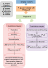
Schematic overview of the experimental approaches applied for the characterization of the venom N-glycomes of Bothrops species by mass spectrometry.
Fig. 2.
Fig. 2.
N-glycans released from glycoproteins present in venoms of the Jararaca Complex (B. alcatraz, B. insularis and B. jararaca).
Fig. 3.
Fig. 3.
Spectra of MSn analysis of structure B11 found in B. alcatraz venom. On the left side is shown the structure of N-glycan B11 and its MS2 spectrum. On the right side is shown the structure after sialic acid dimer and terminal sialic acid loss (ion with m/z 1307.80+2) and its respective MS/MS spectrum. One antenna was chosen for representation of the neutral loss.
Fig. 4.
Fig. 4.
Structural analysis of the two types of sialic acid dimer found in groups 1 and 3 of N-glycans. A, Fragmentation analysis of the sialic acid dimer composed of two NeuAc units and the MS3 spectrum of the ion m/z 759+1 obtained from structure B11 found in B. alcatraz venom. B, Fragmentation analysis of the sialic acid dimer composed of NeuAc and NeuGc units. The MS3 spectrum of the ion m/z 789+1 was obtained from structures B47a and B47b found in B. alcatraz venom.
Fig. 5.
Fig. 5.
Analysis of the isomers B2, B46a and B46b found in the B. jararaca venom. A, Spectrum of the fragments generated from the parent ion (m/z 1292.84+2, [M+Na+] = 2562.44 Da). B, Spectrum obtained from the fragmentation of ion m/z 1105.48+2 (MS3) generated in the spectrum shown in (A). This ion corresponds to the loss of a NeuAc b-ion (neutral loss 375 Da) and suggests the structures B2 and B46a. C, Fragmentation of the ion m/z 1090.48+2 (MS3) generated from the loss of a NeuGc b-ion (neutral loss of 405 Da) and is specific for structure B46b. One antenna was chosen for representation of the neutral loss.
Fig. 6.
Fig. 6.
N-glycans released from glycoproteins present in the venom of B. erythromelas, B. jararacussu, B. moojeni, and B. neuwiedi.
Fig. 7.
Fig. 7.
Antenna fragmentation of structure B32 from B. moojeni venom N-glycome. A, Structure of b-ion NeuAc-2,3-GalNAc-1,4-GlcNAc-1-ene with m/z 888+1 selected for fragmentation (B). C, Structures of b-ion NeuAc-2,3-GalNAc-1-ene and NeuAc-2,6-GalNAc-1-ene (m/z 643+1) with major fragmentations between the monosaccharide units and the cross ring cleavages that allow for the differentiation between these two linkage types. D, Fragmentation of b-ion NeuAc-2,3-GalNAc-1-ene (m/z 643+1) found in the structure B32 from B. moojeni venom N-glycome. The red narrows indicate the presence of 0.4A and 2.4A cross linkage cleavages only observed when NeuAc is linked at the position 3.
Fig. 8.
Fig. 8.
Spectra of MSn analysis of structure B32 found in B. moojeni venom. On the left side is shown the structure of B32 N-glycan and the MS2 spectrum of this structure. On right side is shown the same structure after one sialic acid loss (ion with m/z 1356.40+2) and its respective MS/MS spectrum. One antenna was chosen for representation of the neutral loss.
Fig. 9.
Fig. 9.
N-glycan structures released from glycoproteins present in the venom of B. cotiara.
Fig. 10.
Fig. 10.
Spectra of MSn analysis of structure B22 found in B. cotiara venom. On the left side is shown the structure of B22 N-glycan and the MS2 spectrum of this structure. On the right side is shown the same structure after one sialic acid dimer loss (ion with m/z 1618.12+2) and its respective MS/MS spectrum. One antenna was chosen for representation of the neutral loss.
Fig. 11.
Fig. 11.
MSn analysis performed with structure B22 present only in B. cotiara venom glycoproteins (the structure is shown on the top of the right side). The presence of a monocharged ion of m/z 1112+1 represents the core with the N-bisecting GlcNAc without fucosylated reducing end, which was selected for fragmentation (A). B, Mass spectrum of fragmentation of m/z 852+1 (ion m/z 1112+1 after GlcNAc(1-ene) neutral loss (259 Da). C, Mass spectrum of fragmentation of m/z 648+1 (ion m/z 852+1 after mannose arm loss).
Fig. 12.
Fig. 12.
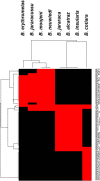
Bothrops venom clustering according to the composition of N-glycans. Graphical visualization of the two hierarchical clusterings of the venom N-glycome characterization. For each venom, a given structure is either present (red) or absent (black).
Fig. 13.
Fig. 13.
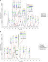
Overlaid LC-MS profiles of the N-glycans present in the venom glycoproteins of the Jararaca Complex (A), B. erythromelas, B. jararacussu, B. moojeni, and B. neuwiedi (B) and B. cotiara (C) and structures that have been assigned in the extracted ion chromatogram.
Fig. 13.
Fig. 13.
Overlaid LC-MS profiles of the N-glycans present in the venom glycoproteins of the Jararaca Complex (A), B. erythromelas, B. jararacussu, B. moojeni, and B. neuwiedi (B) and B. cotiara (C) and structures that have been assigned in the extracted ion chromatogram.

References

    1. Gutiérrez J. M., Williams D., Fan H. W., and Warrell D. A. (2010) Snakebite envenoming from a global perspective: towards an integrated approach. Toxicon 56, 1223–1235 - PubMed
    1. Rägo L., Marroquin A. M. P., Nübling C. M., and Sawyer J. (2015) Treating snake bites—a call for partnership. Lancet 386, 2252 - PubMed
    1. Deshimaru M., Ogawa T., Nakashima K., Nobuhisa I., Chijiwa T., Shimohigashi Y., Fukumaki Y., Niwa M., Yamashina I., Hattori S., et al. (1996) Accelerated evolution of crotalinae snake venom gland serine proteases. FEBS Lett. 397, 83–88 - PubMed
    1. Ohno M., Chijiwa T., Oda-Ueda N., Ogawa T., and Hattori S. (2003) Molecular evolution of myotoxic phospholipases A2 from snake venom. Toxicon 42, 841–854 - PubMed
    1. Fry B. G., Scheib H., van der Weerd L., Young B., McNaughtan J., Ramjan S. F. R., Vidal N., Poelmann R. E., and Norman J. A. (2008) Evolution of an arsenal: structural and functional diversification of the venom system in the advanced snakes (Caenophidia). Mol. Cell. Proteomics 7, 215–246 - PubMed

Publication types