. 2018 May 1;9(3):e00736-17.
doi: 10.1128/mBio.00736-17.

Regulation of Flagellum Biosynthesis in Response to Cell Envelope Stress in Salmonella enterica Serovar Typhimurium

Affiliations

Regulation of Flagellum Biosynthesis in Response to Cell Envelope Stress in Salmonella enterica Serovar Typhimurium

Imke Spöring et al. mBio. .

Abstract

Flagellum-driven motility of Salmonella enterica serovar Typhimurium facilitates host colonization. However, the large extracellular flagellum is also a prime target for the immune system. As consequence, expression of flagella is bistable within a population of Salmonella, resulting in flagellated and nonflagellated subpopulations. This allows the bacteria to maximize fitness in hostile environments. The degenerate EAL domain protein RflP (formerly YdiV) is responsible for the bistable expression of flagella by directing the flagellar master regulatory complex FlhD4C2 with respect to proteolytic degradation. Information concerning the environmental cues controlling expression of rflP and thus about the bistable flagellar biosynthesis remains ambiguous. Here, we demonstrated that RflP responds to cell envelope stress and alterations of outer membrane integrity. Lipopolysaccharide (LPS) truncation mutants of Salmonella Typhimurium exhibited increasing motility defects due to downregulation of flagellar gene expression. Transposon mutagenesis and genetic profiling revealed that σ24 (RpoE) and Rcs phosphorelay-dependent cell envelope stress response systems sense modifications of the lipopolysaccaride, low pH, and activity of the complement system. This subsequently results in activation of RflP expression and degradation of FlhD4C2 via ClpXP. We speculate that the presence of diverse hostile environments inside the host might result in cell envelope damage and would thus trigger the repression of resource-costly and immunogenic flagellum biosynthesis via activation of the cell envelope stress response.IMPORTANCE Pathogenic bacteria such as Salmonella Typhimurium sense and adapt to a multitude of changing and stressful environments during host infection. At the initial stage of gastrointestinal colonization, Salmonella uses flagellum-mediated motility to reach preferred sites of infection. However, the flagellum also constitutes a prime target for the host's immune response. Accordingly, the pathogen needs to determine the spatiotemporal stage of infection and control flagellar biosynthesis in a robust manner. We found that Salmonella uses signals from cell envelope stress-sensing systems to turn off production of flagella. We speculate that downregulation of flagellum synthesis after cell envelope damage in hostile environments aids survival of Salmonella during late stages of infection and provides a means to escape recognition by the immune system.


Figures

FIG 1
FIG 1
Phenotypic characterization of Salmonella LPS mutants regarding motility and flagellation. (A) Schematic representation of LPS structure. Genes encoding the enzymes for particular steps in LPS synthesis were deleted, resulting in the depicted LPS phenotype. (Adapted from reference with permission of the publisher.) (B) Swimming motility of Wt and LPS mutant bacteria assessed on semisolid agar after 4 h of incubation at 37°C. The ΔflhDC mutant strain served as a negative control. Bars represent means + standard errors of the means of results from 2 individual experiments (n = 10). (C) Scanning electron microscopy of Wt Salmonella and the LPS ΔrfaL, ΔrfaG, and ΔrfaD mutants. (D) Western blot of FliC protein levels of Wt and LPS mutants in whole-cell extract (FliC expression; upper panel) and supernatant (SN) (FliC secretion; lower panel). Protein levels were monitored by SDS-PAGE and immunoblotting. The protein DnaK served as an intracellular control.
FIG 2
FIG 2
Analysis of flagellar gene expression in the LPS mutants. (A) Schematic of the hierarchical flagellar gene regulation cascade. The FlhDC flagellar master regulator complex is transcribed from a class 1 promoter. FlhD4C2 induces expression of class 2 promoter genes (e.g., fliL). After completion of the flagellar hook basal body complex, transcription of class 3 promoter genes (e.g., fljB) commences. (B) Relative flhC (class 1), flgE (class 2), and fliC (class 3) gene expression levels of the LPS mutant strains ΔrfaL, ΔrfaG, and ΔrfaD compared to Wt Salmonella. Bars represent means + standard errors of the means of results from 2 individual experiments (n = 6). (C) Degradation assay of FlhC-FLAG protein levels over 60 min. Synthesis was stopped by treatment with spectinomycin and chloramphenicol. A Western blot of FlhC-FLAG protein levels of Wt and mutants in whole-cell extract is shown. Protein levels were monitored by SDS-PAGE and immunoblotting. The protein DnaK served as an intracellular control.
FIG 3
FIG 3
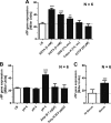
The motility defect of LPS mutants can be mimicked by EDTA supplementation. (A) Change in NPN uptake as an indicator of outer membrane instability in the ΔrfaL, ΔrfaG, and ΔrfaD LPS mutants. Bars represent means + standard errors of the means of results from 1 individual experiment (n = 4). (B) The swimming motility of Wt bacteria was assessed on semisolid agar supplemented with 0, 0.01, 0.1, 1, and 5 mM EDTA and compared to that of ΔrfaG LPS mutant bacteria after 4 h of incubation at 37°C. Bars represent means + standard errors of the means of results from 2 individual experiments (n = 6). (C) Scanning electron microscopy of Wt Salmonella bacteria grown overnight in LB or in LB supplemented with 1 mM EDTA. (D) Relative flhC (class 1), flgE (class 2), and fliC (class 3) gene expression levels of the Wt Salmonella bacteria. The bacteria were grown for 1.5 h in LB. A null experiment was performed or 1 mM EDTA was added, and bacteria were cultured for ~1.5 h prior to gene expression measurement. Bars represent means + standard errors of the means of results from 2 individual experiments (n = 6).
FIG 4
FIG 4
Analysis of flagellar gene expression in the LPS mutants. (A) Schematic model of the stress-mediated downregulation of motility and assumption for design of the transposon screen. The screen is based on the use of the fliL::lac (class 2) reporter gene to screen for mutants that reestablish class 2 gene expression in a ΔrfaG mutant. (B) Schematic depiction of transposon insertion sites found in the screen. (C) Swimming motility of the Wt strain, a ΔrflP mutant, and ΔrfaG and ΔrfaD LPS mutants in the absence and presence of rflP assessed on semisolid agar after 4 h of incubation at 37°C. Bars represent means + standard errors of the means of results from 2 individual experiments (n = 10). (D) Scanning electron microscopy of LPS mutant strains ΔrfaG and ΔrfaD in the absence and presence of rflP. (E) Western blot of FliC protein levels of Wt and LPS mutant strains ΔrfaG and ΔrfaD in the absence or presence of rflP in whole-cell extract (FliC expression; upper panel) and supernatant (SN) (FliC secretion; lower panel). Protein levels were monitored by SDS-PAGE and immunoblotting. The protein DnaK served as an intracellular control. **, P ≤ 0.01; ***, P ≤ 0.001.
FIG 5
FIG 5
Analysis of rflP gene expression in the LPS mutants and upon EDTA addition. (A) Relative rflP gene expression levels analyzed by reverse transcription and quantitative real-time PCR (RT-qPCR) of LPS mutant strains ΔrfaG and ΔrfaD in the absence and presence of rflP compared to Wt Salmonella. Bars represent means + standard errors of the means of results from 3 individual experiments (n = 6). (B) Relative rflP gene expression levels analyzed by RT-qPCR of LPS mutant strains ΔrfaG and ΔrfaD and their complemented strains using the chromosomally integrated PBAD system (ΔrfaG rfaG = ΔaraBAD::rfaG ΔrfaG::aph [EM4410]; ΔrfaD rfaD+ = ΔaraBAD::rfaD ΔrfaD::aph [EM4411]). Bars represent means + standard errors of the means of results from 2 individual experiments (n = 4). (C) Relative rflP gene expression determined by qRT-PCR in LPS mutant strain ΔrfaG compared to Wt Salmonella. The bacteria were grown for 1.5 h in LB. Null experiments were performed or 1 or 5 mM EDTA was added, and bacteria were cultured for ~1.5 h prior to gene expression measurement. Bars represent means + standard errors of the means of results from 2 individual experiments (n = 4). (D) rflP-lac fusion expression in mutant Δspi-1, mutant Δspi-2, and a Δspi-1 Δspi-2 double mutant with and without EDTA. The bacteria were grown for 1.5 h in LB. EDTA was added, and bacteria were cultured for ~1.5 h prior to gene expression measurement by β-galactosidase assay. Bars represent means + standard errors of the means of results from 2 individual experiments (n = 6). *, P ≤ 0.05; **, P ≤ 0.01.
FIG 6
FIG 6
Analysis of rflP gene expression upon addition of various envelope stressing conditions. (A) Chemical membrane stressing agents: EDTA, EGTA, SDS, Triton, and CCCP. (B) Changes of the medium conditions: pH and the antibiotic ampicillin (Amp) and the antimicrobial peptide polymyxin B (Poly B). (C) Human serum containing the complement system. The bacteria were grown for 1.5 h in LB. The stressors were added, and bacteria were cultured for ~1.5 h prior to gene expression measurement by β-galactosidase assay. Bars represent means + standard errors of the means of results from 2 individual experiments (n ≥ 4). **, P ≤ 0.01; ***, P ≤ 0.001.
FIG 7
FIG 7
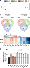
Transcriptome analyses of the LPS mutants reveals involvement of stress sigma factor σ24 in activation of rflP. (A) Principal-component analysis of the transcriptomes of LPS mutants ΔrfaL, ΔrfaG, and ΔrfaD. Colored circles depict the two biological replicates of each mutant. (B) Venn diagram of the LPS mutant transcriptomes. (C) Averaged log2-fold change (log2FC) of expression levels of selected genes of interest in the ΔrfaG and ΔrfaD mutants, including the following: (i) genes involved in transposon mutagenesis (nlpC, rflP, clpX, atp operon); (ii) genes encoding stress response sigma factors (rpoH, rpoE); (iii) genes encoding Rst (rstAB) and Cpx (cpxPR) signal transduction systems; (iv) genes corresponding to an unknown putative two-component system (STM4310-STM4315). (D) rflP expression in ΔrpoE, ΔrcsBDC, ΔyjbE, ΔrstA, ΔrstB, ΔhtrA, ΔcpxAR ΔcpxPcpxARP), and ΔcpxP mutants. The LB medium was supplemented with 1 mM EDTA to induce rflP expression in response to cell envelope stress. Salmonella Wt bacteria grown in LB served as controls. The bacteria were grown for 1.5 h in LB. EDTA (1 mM) was added, and bacteria were cultured for ~1.5 h prior to gene expression measurement. Bars represent means + standard errors of the means of results from 2 individual experiments (n = 6).
FIG 8
FIG 8
Influence of the Rcs pathway on the motility defect in the LPS mutants. (A) Swimming motility of the Wt strain, ΔrfaG mutants, and ΔrfaG Δrcs double mutants assessed on semisolid agar after 4 h of incubation at 37°C. Bars represent means + standard errors of the means of results from 2 individual experiments (n = 4). (B) rflP-lac expression either in an RcsB mutant background that mimics phosphorylation (rcsBD56E—rcsBon) or in a mutant that is unable to be phosphorylated (rcsBD56N—rcsBoff) upon addition of 1 mM EDTA. Salmonella Wt bacteria grown in LB served as controls. The bacteria were grown for 1.5 h in LB. EDTA (1 mM) was added, and bacteria were cultured for ~1.5 h prior to gene expression measurement. Bars represent means + standard errors of the means of results from 2 individual experiments (n = 6).
FIG 9
FIG 9
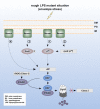
Model of envelope stress-mediated downregulation of flagellar gene expression. Cell envelope stress signals are sensed in the LPS mutant background by various sensory systems located in the membrane. The signal may be transmitted (i) directly via the alternative cell envelope stress sigma factor σ24, which is encoded by the rpoE gene, (ii) indirectly by enhancing transcription of rpoE, (iii) via the Rcs signaling pathway by phosphorylating RcsB, or (iv) via an unknown sensor and signaling pathway. RflP expression is then increased directly by σ24 via the activity of a σ24 binding sequence in the promoter region of rflP, by direct or indirect induction by RcsB-P, or by other factors. Increased levels of RflP protein led to degradation of the flagellar master regulator protein complex FlhD4C2 via ClpXP protease and subsequent downregulation of flagellar synthesis. Genes are indicated by gray boxes, and gene names are in italics.

References

    1. Berg HC, Anderson RA. 1973. Bacteria swim by rotating their flagellar filaments. Nature 245:380–382. doi:10.1038/245380a0. - DOI - PubMed
    1. Renault TT, Abraham AO, Bergmiller T, Paradis G, Rainville S, Charpentier E, Guet CC, Tu Y, Namba K, Keener JP, Minamino T, Erhardt M. 2017. Bacterial flagella grow through an injection-diffusion mechanism. Elife 6:e23136. doi:10.7554/eLife.23136. - DOI - PMC - PubMed
    1. Chevance FF, Hughes KT. 2008. Coordinating assembly of a bacterial macromolecular machine. Nat Rev Microbiol 6:455–465. doi:10.1038/nrmicro1887. - DOI - PMC - PubMed
    1. Macnab RM. 2003. How bacteria assemble flagella. Annu Rev Microbiol 57:77–100. doi:10.1146/annurev.micro.57.030502.090832. - DOI - PubMed
    1. Soutourina OA, Bertin PN. 2003. Regulation cascade of flagellar expression in Gram-negative bacteria. FEMS Microbiol Rev 27:505–523. doi:10.1016/S0168-6445(03)00064-0. - DOI - PubMed

Publication types

MeSH terms

Substances