. 2018 Jun;177(2):713-727.
doi: 10.1104/pp.18.00017. Epub 2018 May 1.

OsMADS6 Controls Flower Development by Activating Rice FACTOR OF DNA METHYLATION LIKE1

Affiliations

OsMADS6 Controls Flower Development by Activating Rice FACTOR OF DNA METHYLATION LIKE1

Juhong Tao et al. Plant Physiol. 2018 Jun.

Abstract

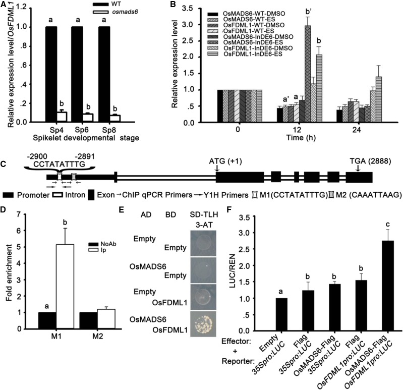
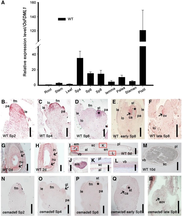
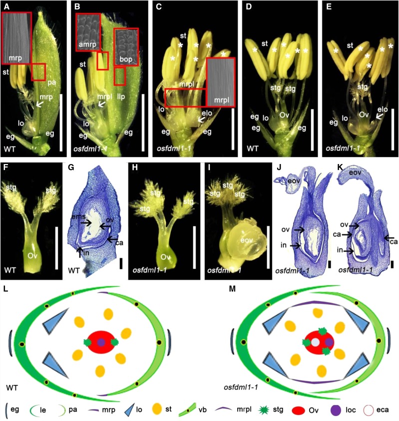
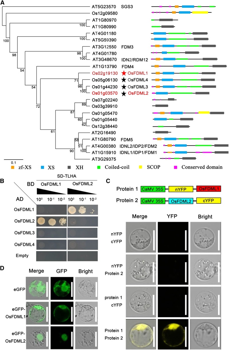
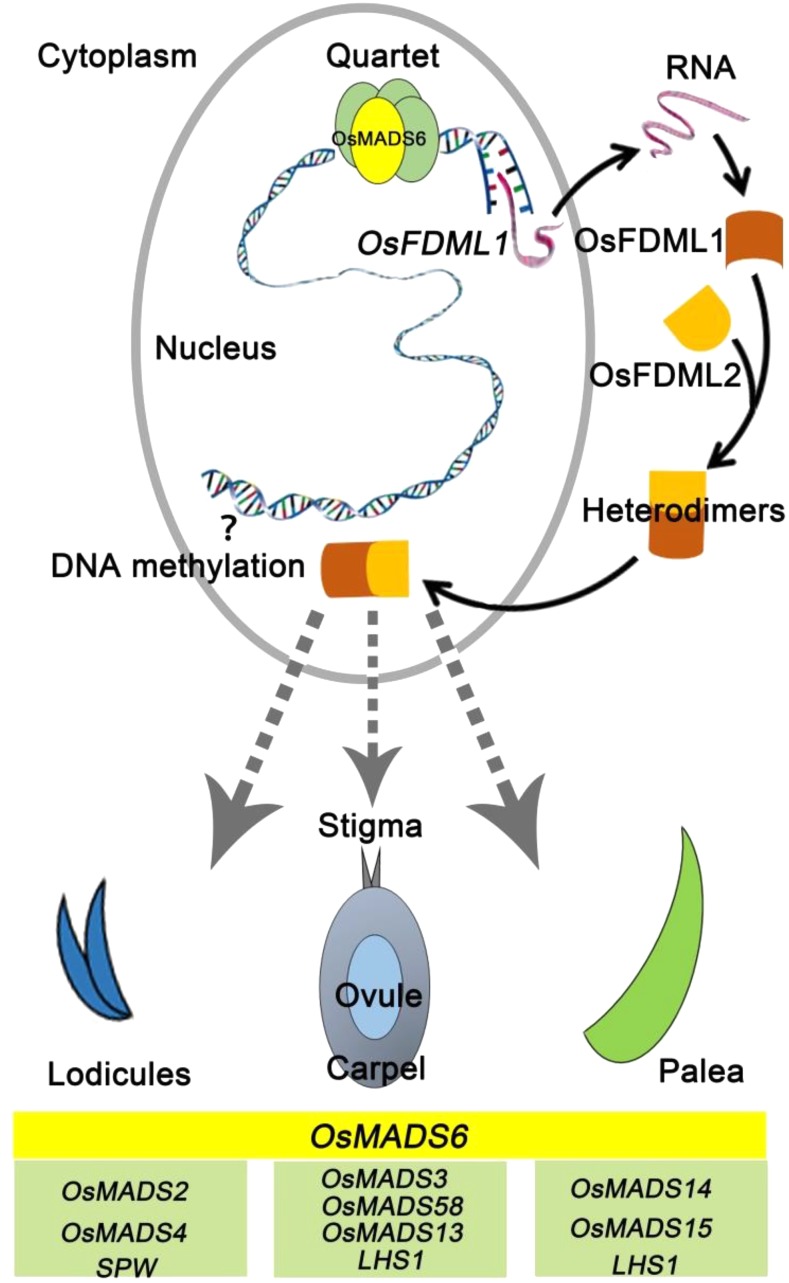
OsMADS6, an ancient AGAMOUS-LIKE6 (AGL6)-like gene, has essential functions in specifying floral organ and meristem identity in rice (Oryza sativa). However, how AGL6 genes control flower development remains largely unknown. In this study, we report that OsMADS6 directly targets FACTOR OF DNA METHYLATION LIKE 1 (OsFDML1), a rice homolog of the SUPPRESSOR OF GENE SILENCING3-like gene FACTOR OF DNA METHYLATION 1 (FDM1) from Arabidopsis (Arabidopsis thaliana). Arabidopsis FDM1 is involved in RNA-directed DNA methylation and OsFDML1 regulates flower development. The expression of OsFDML1 overlaps with that of OsMADS6 in the palea primordia and the ovule, and OsMADS6 directly promotes OsFDML1 expression through binding to regions containing putative CArG motifs within the OsFDML1 promoter during rice spikelet development. Consistent with the phenotypes of osmads6 mutants, the osfdml1 mutants showed floral defects, including altered palea identity with lemma-like shape containing no marginal region of palea, increased numbers of stigmas and fused carpels, and meristem indeterminacy. Moreover, transgenic plants overexpressing OsFDML1 displayed floral defects, such as abnormal paleae. Phylogenetic analysis showed that OsFDML1 homologs exist only in terrestrial plants. In addition, protein-protein interaction assays showed that OsFDML1 interacts with its close paralog OsFDML2, similar to the activity of OsFDML1 homologs in Arabidopsis. These results provide insight into how the ancient AGL6 gene regulates floral development.


Figures

Figure 1.
Figure 1.
OsMADS6 directly activates the expression of OsFDML1 in vivo during rice spikelet development. A, The expression of OsFDML1 decreased in osmads6-1 at spikelet developmental stages Sp4, Sp6, and Sp8. Spikelets at stages Sp4, Sp6, and Sp8 were collected from inflorescences of lengths 2, 2 to 3, and 4 to 7 mm, respectively. B, The expression of OsFDML1 increased with the induced expression of OsMADS6 in the transgenic plants InDE6. C, Diagram of OsFDML1 gene with promoter regions containing the putative CArG motif. D, ChIP-qPCR results showed OsMADS6 can directly bind to OsFDML1 promoter regions with putative CArG motif (M1) using 2- to 8-mm-long young inflorescences. The mock treatment included negative control treated without antibody and 200-bp OsFDML1 promoter region containing an unclassical CArG motif M2 close by to the motif M1. The fold enrichment was calculated relative to the negative control without antibody. E, OsMADS6 bound to the OsFDML1 promoter regions with putative CArG motif (M1) and activated the expression of nutritional reporter gene HIS3 by yeast one-hybrid assay. F, OsMADS6 triggered the expression of OsFDML1pro:LUC with integrating 3795-bp OsFDML1 genome sequence upstream of the ATG before LUC. All data are means ± sd (n ≥ 3). Letters a, b, c, a’, and b’ represent statistical significance, P < 0.05. IP, Immunoprecipitation with specific antibody against OsMADS6; InDE6, transgenic plants containing OsMADS6 fused with XVE; ES, β-estradiol.
Figure 2.
Figure 2.
Expression pattern of OsFDML1 in the wild-type and osmads6-1 young spikelets. A, RT-qPCR analysis of the expression of OsFDML1 in wild-type root, stem, leaf, and spikelets at stages Sp4, Sp6, and early Sp8, and dissected lemma, palea, stamen, and pistil, respectively. Root was collected from young seedlings cultivated 10 d in water. Stem and leaf were collected when the plants changed to reproductive growth phase. Spikelets at stages Sp4, Sp6, and Sp8 were sampled from inflorescences of lengths ∼2, 2 to 3, and 4 to 7 mm, respectively. Lemma, palea, stamen, and pistil were dissected from the spikelets of length ∼6 mm. All data are means ± sd (n ≥ 3). B to F, In situ hybridization experiment showing the expression of OsFDML1 in wild-type young spikelets at stages Sp2, Sp4, Sp6, early Sp8, and late Sp8. Spikelets at stages Sp2, Sp4, Sp6, early Sp8, and late Sp8 were sampled from inflorescences of lengths ∼1 to 1.5, 2, 2 to 3, 4 to 7, and 10 to 20 mm, respectively. Asterisks indicate the palea side stamen in D and E. Arrowheads indicate the carpel primordia in E and the ovule in F. G to M, In situ hybridization experiment showing the expression of OsFDML1 in wild-type ovaries representing 0, 2, 5, and 10 d since pollination. J to L, partial enlargements of I. Arrowheads indicate the integument and nucellus in G and H, respectively. Red rectangles indicate the position of J to L in I. N to R, In situ hybridization experiment showing the expression of OsFDML1 in osmads6-1 spikelets at stages Sp2, Sp4, Sp6, early Sp8, and late Sp8, respectively. Asterisks indicate the palea side stamens in P and Q. Arrowheads indicate the glume-like structure of osmads6-1 in O, carpel primordia of osmads6-1 in Q, and the abnormal ovule of osmads6-1 in R. le, Lemma; pa, palea; gl, glume-like structure; lo, lodicules; st, stamen; fm, floral meristem; ca, carpel; ov, ovule; aov, abnormal ovule; nu, nucellus; in, integument; em, embryo; pl, pericarp; sc, seed capsule; al, aleurone; vb, vascular bundle. Bars = 50 μm in B, C, F, N, O, and R, 100 μm in D, E, J to L, P, and Q, and 500 μm in G to I and M.
Figure 3.
Figure 3.
Phenotypes of the wild-type and osfdml1-1 spikelets. A, Wild-type spikelet (genetic background Hwayoung) with dissected lemma. Arrowhead indicates the MRP structure. The red rectangles indicate the position of scanning electron microscopy and the cell identity of the MRP structure. B, osfdml1-1 spikelets developing a lemma-like palea in whorl 1 and MRP-like structures in whorl 2. Arrowhead indicates the MRP-like structure. The red rectangles indicate the positions of scanning electron microscopy and the cell identity of the BOP and abnormal MRP structures. C, osfdml1-1 spikelet with dissected lemma and the lemma-like palea showing extra lodicules and MRP-like structure in whorl 2. Arrowhead indicates the extra lodicules in the palea side. The red rectangles indicate the positions of scanning electron microscopy and the cell identity of the MRP-like structure. D, Wild-type spikelet with lodicules in lemma side, six stamens, and one pistil with two stigmas. Asterisks indicate the stamens. E, osfdml1-1 spikelets showing extra lodicules in palea side. Arrowhead indicates the extra lodicules in palea side. Asterisks indicate the stamens. F, Wild-type pistil with two stigmas. G, Vertical section of the wild-type ovary. Arrowheads indicate the carpel, integument, ovule, and embryo sac. H, osfdml1-1 pistil with three stigmas. I, osfdml1-1 pistil with three stigmas and an exposed ovule. J and K, Vertical section of the osfdml1-1 ovary. Occasionally two ovaries fused together (K). Arrowheads indicate the carpel, integument, and ovule. L and M, Diagrams of the wild-type and osfdml1-1 spikelets. eg, Empty glum; pa, palea; llp, lemma-like palea; st, stamen; lo, lodicule; elo, extra lodicules; mrp, marginal region of palea; mrpl, mrp-like structure; bop, body of palea; amrp, abnormal mrp structure; Ov, ovary; stg, stigma; ov, ovule; in, integument; ems, embryo sac; ca, carpel; eov, exposed ovule; vb, vascular bundle; loc, locule; eca, extra carpel. Bars = 2 mm in A and B, 1 mm in C to E, 500 μm in F, H, and I, and 100 μm in G, J, and K.
Figure 4.
Figure 4.
Phenotypes of OsFDML1 ectopic-expressed spikelets. A and B, Wild-type spikelet (A) with dissected lemma (B). Arrowhead indicates the MRP structure. The red rectangles indicate the positions of scanning electron microscopy and the cell identity of the BOP and MRP structures. C and D, Phenotype of OsFDML1 overexpressing spikelet (C) with ectopic wider MRP structure (D). Arrowhead indicates the ectopic MRP structure. The red rectangles indicate the position of scanning electron microscopy and the cell identity of the BOP, BOP-like, and MRP structures. E and F, Cross section of the wild-type MRP (E) and the ectopic wider MRP structure of OET6 (F). The vascular bundles are marked by red rectangles. Double-headed arrow indicates the additional BOP-like structure. G, Statistical analysis of abnormal flowers of the wild-type and OET6 inflorescences. All data are means ± sd (n ≥ 5). H, Relative expression level of OsFDML1 of the wild-type and OET6 spikelet. All data are means ± sd (n ≥ 3). Letters a and b represented statistical significance, P < 0.05. eg, Empty glume; le, lemma; pa, palea; mrp, marginal region of palea; emrp, ectopic mrp structure; bop, body of palea; bopl, bop-like structure; lo, lodicule; st, stamen; vb, vascular bundle; avb, additional vascular bundle. Bars = 2 mm in A to D and 100 μm in E and F.
Figure 5.
Figure 5.
OsFDML1 functions as heterodimers with its close paralog OsFDML2. A, The neighbor-joining phylogenetic tree of OsFDML1 with homologs in rice and Arabidopsis. The bootstrap values (%) based on 100 replicates are marked at the branching points. The scale bar indicates the number of nucleotide substitutions per site. OsFDML1 and the interacting protein OsFDML2 are marked in red. The red star indicates OsFDML1. The black stars indicate the other three paralogs OsFDML2, OsFDML3, and OsFDML4 in rice belonging to the same subgroup. B, OsFDML1 interacts with its close paralog OsFDML2 by yeast two-hybrid assay. C, OsFDML1 interacts with OsFDML2 in vivo by BiFC assay in rice protoplast. D, OsFDML1 localizes in nucleus and cytoplasm, and OsFDML2 localizes in nucleus in rice protoplast. Bars = 10 μm in C and D.
Figure 6.
Figure 6.
An updated model for the regulation of palea, lodicule, and pistil development by OsMADS6 via direct activation of OsFDML1. OsFDML1 functions as heterodimers with its close paralog OsFDML2.

References

    1. Agrawal GK, Abe K, Yamazaki M, Miyao A, Hirochika H (2005) Conservation of the E-function for floral organ identity in rice revealed by the analysis of tissue culture-induced loss-of-function mutants of the OsMADS1 gene. Plant Mol Biol 59: 125–135 - PubMed
    1. Ambrose BA, Lerner DR, Ciceri P, Padilla CM, Yanofsky MF, Schmidt RJ (2000) Molecular and genetic analyses of the silky1 gene reveal conservation in floral organ specification between eudicots and monocots. Mol Cell 5: 569–579 - PubMed
    1. Arora R, Agarwal P, Ray S, Singh AK, Singh VP, Tyagi AK, Kapoor S (2007) MADS-box gene family in rice: genome-wide identification, organization and expression profiling during reproductive development and stress. BMC Genomics 8: 242. - PMC - PubMed
    1. Ausin I, Mockler TC, Chory J, Jacobsen SE (2009) IDN1 and IDN2 are required for de novo DNA methylation in Arabidopsis thaliana. Nat Struct Mol Biol 16: 1325–1327 - PMC - PubMed
    1. Ausin I, Greenberg MV, Simanshu DK, Hale CJ, Vashisht AA, Simon SA, Lee TF, Feng S, Española SD, Meyers BC, et al. (2012) INVOLVED IN DE NOVO 2-containing complex involved in RNA-directed DNA methylation in Arabidopsis. Proc Natl Acad Sci USA 109: 8374–8381 - PMC - PubMed

Publication types