Comparative Study
. 2018 May 15;115(20):E4700-E4709.
doi: 10.1073/pnas.1721395115. Epub 2018 May 1.

Comparative genomics of the nonlegume Parasponia reveals insights into evolution of nitrogen-fixing rhizobium symbioses

Affiliations
Comparative Study

Comparative genomics of the nonlegume Parasponia reveals insights into evolution of nitrogen-fixing rhizobium symbioses

Robin van Velzen et al. Proc Natl Acad Sci U S A. .

Abstract

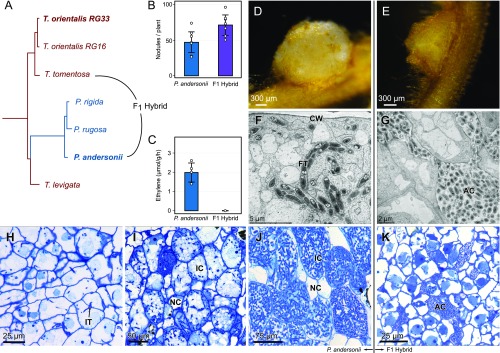
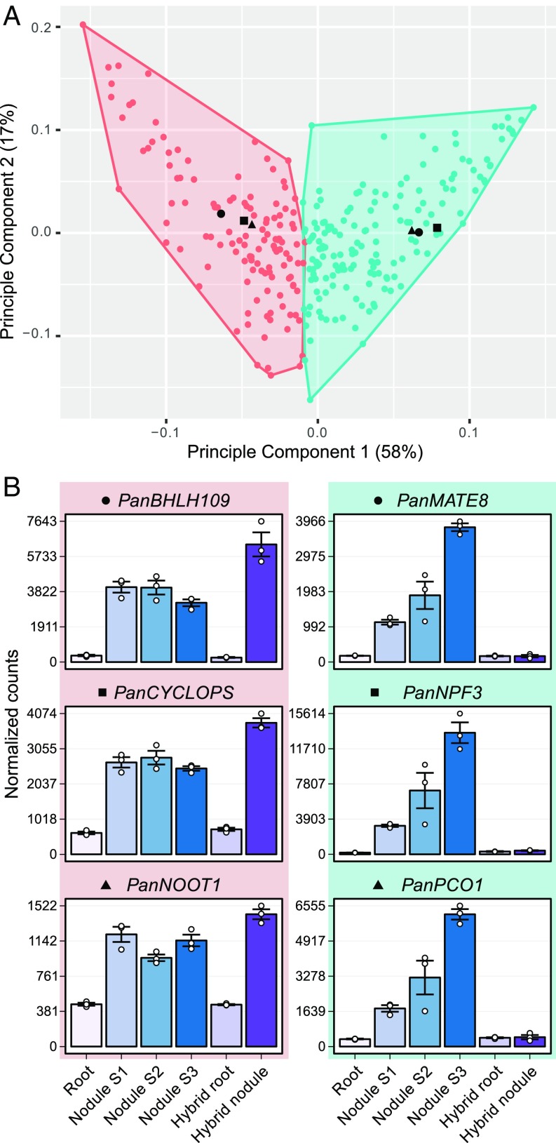
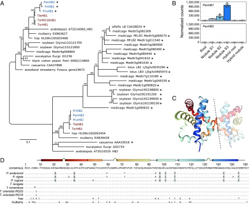
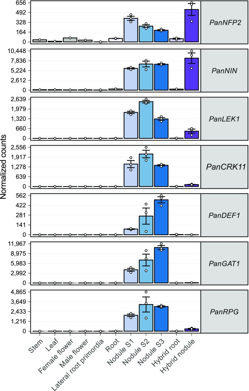
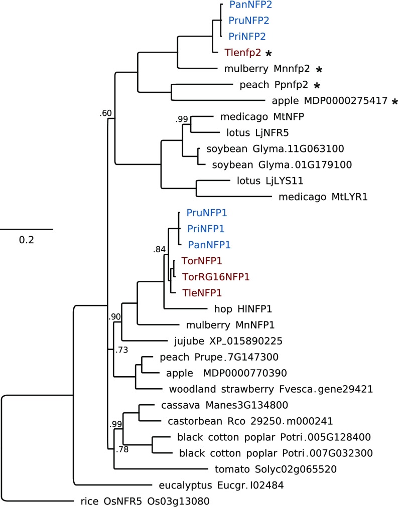
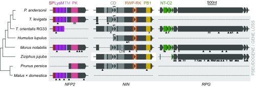
Nodules harboring nitrogen-fixing rhizobia are a well-known trait of legumes, but nodules also occur in other plant lineages, with rhizobia or the actinomycete Frankia as microsymbiont. It is generally assumed that nodulation evolved independently multiple times. However, molecular-genetic support for this hypothesis is lacking, as the genetic changes underlying nodule evolution remain elusive. We conducted genetic and comparative genomics studies by using Parasponia species (Cannabaceae), the only nonlegumes that can establish nitrogen-fixing nodules with rhizobium. Intergeneric crosses between Parasponia andersonii and its nonnodulating relative Trema tomentosa demonstrated that nodule organogenesis, but not intracellular infection, is a dominant genetic trait. Comparative transcriptomics of P. andersonii and the legume Medicago truncatula revealed utilization of at least 290 orthologous symbiosis genes in nodules. Among these are key genes that, in legumes, are essential for nodulation, including NODULE INCEPTION (NIN) and RHIZOBIUM-DIRECTED POLAR GROWTH (RPG). Comparative analysis of genomes from three Parasponia species and related nonnodulating plant species show evidence of parallel loss in nonnodulating species of putative orthologs of NIN, RPG, and NOD FACTOR PERCEPTION Parallel loss of these symbiosis genes indicates that these nonnodulating lineages lost the potential to nodulate. Taken together, our results challenge the view that nodulation evolved in parallel and raises the possibility that nodulation originated ∼100 Mya in a common ancestor of all nodulating plant species, but was subsequently lost in many descendant lineages. This will have profound implications for translational approaches aimed at engineering nitrogen-fixing nodules in crop plants.

Keywords: biological nitrogen fixation; comparative genomics; copy number variation; evolution; symbiosis.


Conflict of interest statement

The authors declare no conflict of interest.

Figures

Fig. 1.
Fig. 1.
Nodulation phenotype of P. andersonii and interspecific P. andersonii × T. tomentosa F1 hybrid plants. (A) Phylogenetic reconstruction based on whole chloroplast of Parasponia and Trema. The Parasponia lineage (blue) is embedded in the Trema genus (red). Species selected for interspecific crosses are indicated and species used for reference genome assembly are in bold. All nodes had a posterior probability of 1. (B) Mean number of nodules on roots of P. andersonii and F1 hybrid plants (n = 7). (C) Mean nitrogenase activity in acetylene reductase assay of P. andersonii and F1 hybrid nodules (n = 4). Bar-plot error bars indicate SDs; dots represent individual measurements. (D) P. andersonii nodule. (E) F1 hybrid nodule. (F and G) Ultrastructure of nodule tissue of P. andersoni (F) and F1 hybrid (G). Note the intracellular fixation thread (FT) in the cell of P. andersonii in comparison with the extracellular, apoplastic colonies of rhizobia (AC) in the F1 hybrid nodule. (HJ) Light-microscopy images of P. andersonii nodules in three subsequent developmental stages. (H) Stage 1: initial infection threads (IT) enter the host cells. (I) Stage 2: progression of rhizobium infection in nodule host cell. (J) Stage 3: nodule cells completely filled with fixation threads. Note difference in size between the infected (IC) and noninfected cells (NC). (K) Light-microscopy image of F1 hybrid nodule cells. Note rhizobium colonies in apoplast, surrounding the host cells (AC). Nodules have been analyzed 6 wk post inoculation with M. plurifarium BOR2. CW, cell wall.
Fig. 2.
Fig. 2.
Clustering of commonly utilized symbiosis genes based on expression profile in P. andersonii. (A) Principal component analysis plot of the expression profile of 290 commonly utilized symbiosis genes in 18 transcriptome samples: P. andersonii roots and nodules (stage 1–3) and hybrid roots and nodules (line H9). All samples have three biological replicates. The first two components are shown, representing 75% of the variation in all samples. Colors indicate clusters (k-means clustering using Pearson correlation as distance measure, k = 2) of genes with similar expression patterns. The three genes with the highest Pearson correlation to the cluster centroids are indicated as black dots, triangles, and squares, and their expression profiles are given in B. Cluster 1 (pink) represents genes related to nodule organogenesis: these genes are up-regulated in P. andersonii and hybrid nodules. Cluster 2 (green) represents genes related to infection and fixation: these genes are highly up-regulated in P. andersonii nodules but do not respond in the hybrid nodule. PanBHLH109, BASIC HELIX–LOOP–HELIX DOMAIN CONTAINING PROTEIN 109; PanMATE8, MULTI ANTIMICROBIAL EXTRUSION PROTEIN 8; PanNOOT1, NODULE ROOT 1; PanNPF3, NITRATE/PEPTIDE TRANSPORTER FAMILY 3; PanPCO1, PLANT CYSTEINE OXIDASE 1.
Fig. 3.
Fig. 3.
Parasponia-specific adaptations in class 1 hemoglobin protein HB1. (A) Phylogenetic reconstruction of class 1 (OG0010523) and class 2 hemoglobins (OG0002188). Symbiotic hemoglobins are marked with an asterisk; legumes and the actinorhizal plant casuarina have recruited class 2 hemoglobins for balancing oxygen levels in their nodules. Conversely, Parasponia has recruited a class 1 hemoglobin PanHB1, confirming parallel evolution of symbiotic oxygen transport in this lineage: M. truncatula (Medtr), G. max (Glyma), P. trichocarpa (Potri), F. vesca (Fvesca), E. grandis (Eugr), A. thaliana (AT). Node values indicate posterior probabilities below 1; scale bar represents substitutions per site. Parasponia marked in blue, Trema in red. (B) Expression profile of PanHB1 and PanHB2 in P. andersonii roots, stage 1–3 nodules, and P. andersonii × T. tomentosa F1 hybrid roots and nodules (line H9). Expression is given in DESeq2-normalized read counts; error bars represent SE of three biological replicates and dots represent individual expression levels. (C) Crystal structure of the asymmetric dimer of PanHB1 as deduced by Kakar et al. (48). Dashed line separates the two units. (D) Protein sequence alignment of class 1 hemoglobins from Parasponia spp.,Trema spp., hop (H. lupulus), and mulberry (M. notabilis). Only amino acids that differ from the consensus are drawn. A linear model of the crystal structure showing α-helices and turns is depicted above the consensus sequence. There are seven amino acids (marked gray) that consistently differ between all Parasponia and all other sampled species: Ala(21), Gln(35), Asp(97), Ile(101), Thr(108), Val(144), and Phe(155). These differences therefore correlate with the functional divergence between P. andersonii PanHB1 and T. tomentosa TtoHB1 (47, 48).
Fig. 4.
Fig. 4.
Expression profile of P. andersonii symbiosis genes that are lost in Trema species. Expression of symbiosis genes in P. andersonii stem, leaf, female and male flowers, lateral root primordia, roots, and three nodule stages (S1–S3), and in F1 hybrid roots and nodules (line H9). Expression is given in DESeq2-normalized read counts; error bars represent SE of three biological replicates for lateral root primordia, root, and nodule samples. Dots represent individual expression levels. PanCRK11, CYSTEINE-RICH RECEPTOR KINASE 11; PanDEF1, DEFENSIN 1; PanLEK1, LECTIN RECEPTOR KINASE 1; PanNFP2, NOD FACTOR PERCEPTION 2; PanNIN, NODULE INCEPTION; PanRPG, RHIZOBIUM DIRECTED POLAR GROWTH.
Fig. 5.
Fig. 5.
Parasponia NFP2 are putative orthologs of legume LCO receptors MtNFP/LjNFR5. Phylogenetic reconstruction of the NFP/NFR5 orthogroup based on kinase domain. Protein sequences deduced from pseudogenes are marked with an asterisk. Included species are P. andersonii (Pan), Parasponia rigida (Pri), Parasponia rugosa (Pru), T. orientalis RG33 (Tor), T. orientalis RG16 (TorRG16), T. levigata (Tle), medicago (M. truncatula, Mt), lotus (L. japonicus, Lj), soybean (G. max, Glyma), peach (P. persica, Ppe), woodland strawberry (F. vesca, Fvesca), black cotton poplar (P. trichocarpa, Potri), eucalyptus (E. grandis, Eugr), jujube (Z. jujuba), apple (M. × domestica), mulberry (M. notabilis), hops (H. lupulus), cassava (Manihot esculenta), rice (O. sativa), tomato (S. lycopersicum), and castor bean (Ricinus communis). Node numbers indicate posterior probabilities below 1; scale bar represents substitutions per site. Parasponia proteins are marked in blue, Trema in red.
Fig. 6.
Fig. 6.
Parallel loss of symbiosis genes in nonnodulating Rosales species. Pseudogenization or loss of NFP2, NIN, and RPG in two phylogenetically independent Trema lineages, H. lupulus (hop), M. notabilis (mulberry), Z. jujuba (jujube), P. persica (peach), and M. × domestica (apple). In H. lupulus, NIN is pseudogenized, whereas NFP2 and RPG were not found (this may be because of the low N50 of the publicly available assembly). In Z. jujuba, NFP2 is lost and RPG is pseudogenized, but NIN is intact. In F. vesca, all three genes are lost (not shown). Introns are indicated but not scaled. Triangles indicate frame shifts; “X” indicates premature stop codons; “LTR” indicates LTR retrotransposon insertion (not scaled); arrows indicate alternative transcriptional start site in NIN. CD, 4 conserved domains (gray); LysM, 3 Lysin motif domains (magenta); NT-C2, N-terminal C2 domain (green); PB1, Phox and Bem1 domain (yellow); PK, protein kinase (pink); RWP-RK, conserved amino acid domain (orange); SP, signal peptide (red); TM, transmembrane domain (lilac).

References

    1. Soltis DE, et al. Chloroplast gene sequence data suggest a single origin of the predisposition for symbiotic nitrogen fixation in angiosperms. Proc Natl Acad Sci USA. 1995;92:2647–2651. - PMC - PubMed
    1. Udvardi M, Poole PS. Transport and metabolism in legume-rhizobia symbioses. Annu Rev Plant Biol. 2013;64:781–805. - PubMed
    1. Ott T, et al. Symbiotic leghemoglobins are crucial for nitrogen fixation in legume root nodules but not for general plant growth and development. Curr Biol. 2005;15:531–535. - PubMed
    1. Burrill TJ, Hansen R. Is symbiosis possible between legume bacteria and non-legume plants? Bull Univ Ill Agric Expt Stn. 1917;202:115–181.
    1. Stokstad E. The nitrogen fix. Science. 2016;353:1225–1227. - PubMed

Publication types