Review
. 2018 Jul 1;98(3):1143-1167.
doi: 10.1152/physrev.00034.2016.

(Re)generating Human Beta Cells: Status, Pitfalls, and Perspectives

Affiliations
Review

(Re)generating Human Beta Cells: Status, Pitfalls, and Perspectives

Luc Baeyens et al. Physiol Rev. .

Abstract

Diabetes mellitus results from disturbed glucose homeostasis due to an absolute (type 1) or relative (type 2) deficiency of insulin, a peptide hormone almost exclusively produced by the beta cells of the endocrine pancreas in a tightly regulated manner. Current therapy only delays disease progression through insulin injection and/or oral medications that increase insulin secretion or sensitivity, decrease hepatic glucose production, or promote glucosuria. These drugs have turned diabetes into a chronic disease as they do not solve the underlying beta cell defects or entirely prevent the long-term complications of hyperglycemia. Beta cell replacement through islet transplantation is a more physiological therapeutic alternative but is severely hampered by donor shortage and immune rejection. A curative strategy should combine newer approaches to immunomodulation with beta cell replacement. Success of this approach depends on the development of practical methods for generating beta cells, either in vitro or in situ through beta cell replication or beta cell differentiation. This review provides an overview of human beta cell generation.


Figures

FIGURE 1.
FIGURE 1.
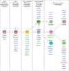
Schematic representation of human pancreas development. Top panel: human pancreas development is shown in relation to the Carnegie classification system (CS; numbers 9 to 23). Embryos are drawn proportional to the UNSW Human Embryo Resource. Middle panel: pancreas morphogenesis is not depicted proportionally but is intended to frame the key developmental steps (bottom panel) in the development of the organ. The human embryonic pancreas evolves as one dorsal and two ventral buds from the primitive foregut (yellow tube) at CS12–13. Prior to budding, the dorsal prepancreatic endoderm is in contact with the notochord (red line). After regression of the left ventral bud, the right ventral bud fuses with the dorsal bud around CS22–23. Bottom panel: key developmental steps include branching morphogenesis (green, tip cells; orange, trunk cells) (A), the first appearance of NEUROG3+ cells (pink) (B), formation of the initial hormone+ cells (green) (C), and establishment of lineage-committed monohormonal endocrine cells (green, red, brown, purple) and formation of the islet cytoarchitecture (D).
FIGURE 2.
FIGURE 2.
Overview of transcription factor expression during human beta cell development. Schematic overview of the signaling cascade governing human pancreas development including currently known transcription factors responsible for cell type fate determination. Transcription factors depicted in green are associated with the development of monogenic diabetes; in red are the transcription factors known to cause MODY.
FIGURE 3.
FIGURE 3.
Nonpancreatic cell sources for beta cell generation. Illustration of the potential use of nonpancreatic cell types to generate new beta(-like) cells in vitro based on hES/hIPS directed differentiation or postnatal donor cell reprogramming. The cells of origin depicted represent healthy donor cells. Functional beta(-like) cells could be reintroduced to replenish the beta cell pool lost in diabetes patients.
FIGURE 4.
FIGURE 4.
Strategies to increase human beta cell numbers in vitro from pancreatic cell types. Schematic overview of the strategies used to generate new beta(-like) cells in vitro using cell types of pancreatic origin. Black arrows represent strategies that have been documented; red arrow depicts a potential strategy that has yet to be shown using human cells.

References

    1. Abouna S, Old RW, Pelengaris S, Epstein D, Ifandi V, Sweeney I, Khan M. Non-β-cell progenitors of β-cells in pregnant mice. Organogenesis 6: 125–133, 2010. doi:10.4161/org.6.2.10374. - DOI - PMC - PubMed
    1. Al-Hasani K, Pfeifer A, Courtney M, Ben-Othman N, Gjernes E, Vieira A, Druelle N, Avolio F, Ravassard P, Leuckx G, Lacas-Gervais S, Ambrosetti D, Benizri E, Hecksher-Sorensen J, Gounon P, Ferrer J, Gradwohl G, Heimberg H, Mansouri A, Collombat P. Adult duct-lining cells can reprogram into β-like cells able to counter repeated cycles of toxin-induced diabetes. Dev Cell 26: 86–100, 2013. doi:10.1016/j.devcel.2013.05.018. - DOI - PubMed
    1. Alonso LC, Yokoe T, Zhang P, Scott DK, Kim SK, O’Donnell CP, Garcia-Ocaña A. Glucose infusion in mice: a new model to induce beta-cell replication. Diabetes 56: 1792–1801, 2007. doi:10.2337/db06-1513. - DOI - PMC - PubMed
    1. Alvarez-Perez JC, Ernst S, Demirci C, Casinelli GP, Mellado-Gil JM, Rausell-Palamos F, Vasavada RC, Garcia-Ocaña A. Hepatocyte growth factor/c-Met signaling is required for β-cell regeneration. Diabetes 63: 216–223, 2014. doi:10.2337/db13-0333. - DOI - PMC - PubMed
    1. Amaral ME, Cunha DA, Anhê GF, Ueno M, Carneiro EM, Velloso LA, Bordin S, Boschero AC. Participation of prolactin receptors and phosphatidylinositol 3-kinase and MAP kinase pathways in the increase in pancreatic islet mass and sensitivity to glucose during pregnancy. J Endocrinol 183: 469–476, 2004. doi:10.1677/joe.1.05547. - DOI - PubMed

Publication types