Review
. 2018 Jul 1;98(3):1241-1334.
doi: 10.1152/physrev.00043.2017.

Gestational Hypoxia and Developmental Plasticity

Affiliations
Review

Gestational Hypoxia and Developmental Plasticity

Charles A Ducsay et al. Physiol Rev. .

Abstract

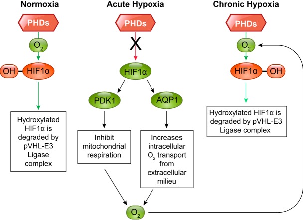
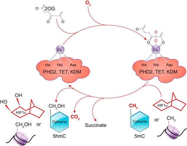
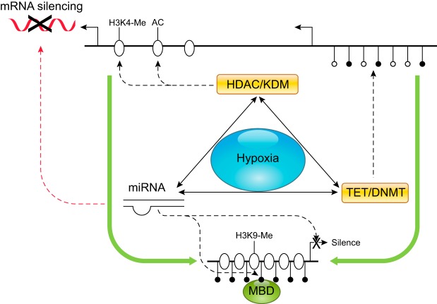
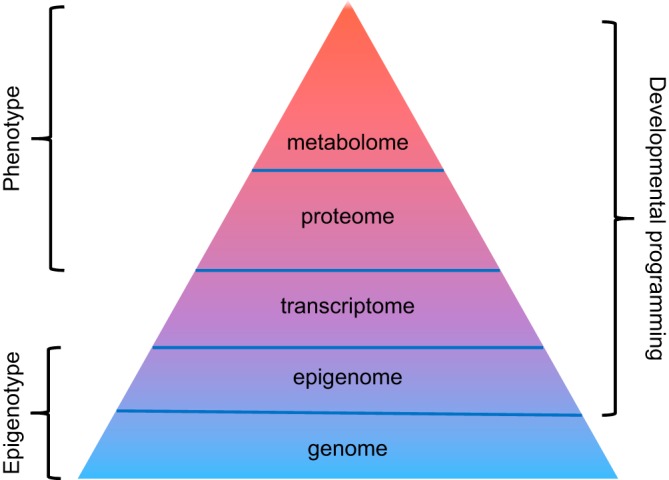
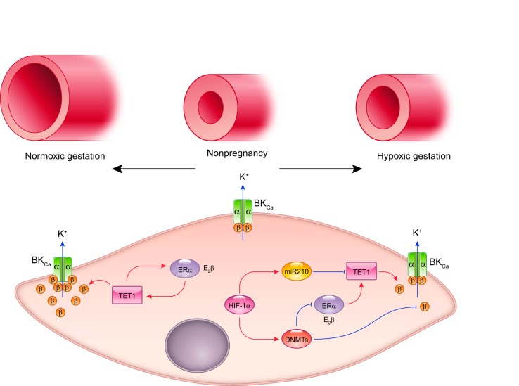
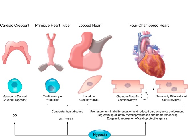
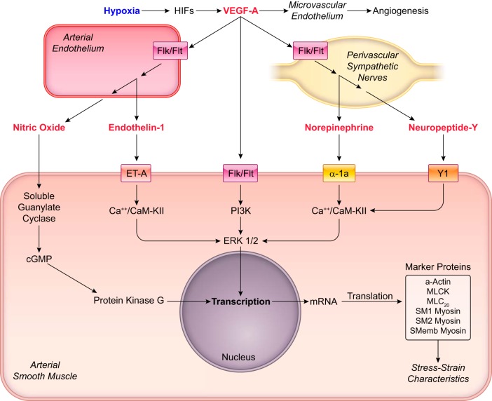
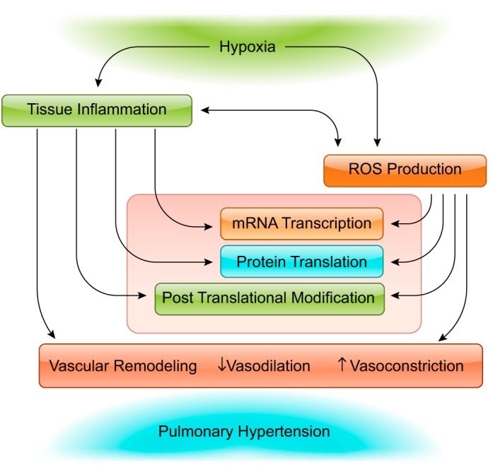
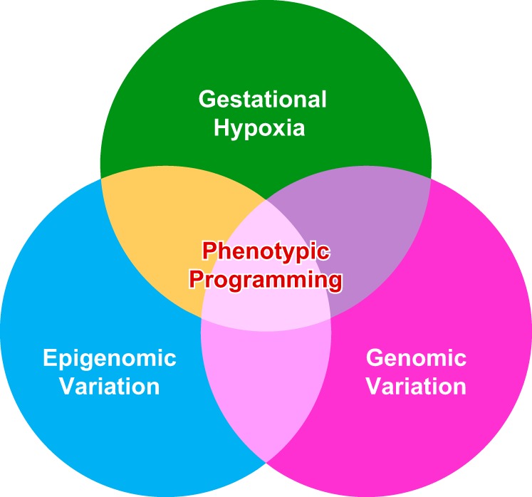
Hypoxia is one of the most common and severe challenges to the maintenance of homeostasis. Oxygen sensing is a property of all tissues, and the response to hypoxia is multidimensional involving complicated intracellular networks concerned with the transduction of hypoxia-induced responses. Of all the stresses to which the fetus and newborn infant are subjected, perhaps the most important and clinically relevant is that of hypoxia. Hypoxia during gestation impacts both the mother and fetal development through interactions with an individual's genetic traits acquired over multiple generations by natural selection and changes in gene expression patterns by altering the epigenetic code. Changes in the epigenome determine "genomic plasticity," i.e., the ability of genes to be differentially expressed according to environmental cues. The genomic plasticity defined by epigenomic mechanisms including DNA methylation, histone modifications, and noncoding RNAs during development is the mechanistic substrate for phenotypic programming that determines physiological response and risk for healthy or deleterious outcomes. This review explores the impact of gestational hypoxia on maternal health and fetal development, and epigenetic mechanisms of developmental plasticity with emphasis on the uteroplacental circulation, heart development, cerebral circulation, pulmonary development, and the hypothalamic-pituitary-adrenal axis and adipose tissue. The complex molecular and epigenetic interactions that may impact an individual's physiology and developmental programming of health and disease later in life are discussed.


Figures

FIGURE 1.
FIGURE 1.
Hypoxia and developmental origins of health and disease. Gestational hypoxia is associated with women residing at high altitude, for whom having preeclampsia and placental insufficiency imposes a significant challenge to both the mother and the developing fetus. To cope with such an adverse environmental condition, the mother and her fetus as well as the placenta undergo significant changes to achieve a certain degree of adaptation to ensure reproductive success and to sustain life before birth. Notably, the expression patterns of suites of genes encoding receptors, ion channels, enzymes, and other signal proteins are altered via epigenetic modifications. However, certain trade-offs occur along with this adaptation and result in permanent changes in phenotypic plasticity and cell/tissue function, leading to increased risk of disease later in life.
FIGURE 2.
FIGURE 2.
Illustration of the interplay of external factors on developmental plasticity and the downstream effects on general and system specific adaptations in the developing fetus. Also shown are the known disorders associated with such interactions.
FIGURE 3.
FIGURE 3.
Illustration of the effect of various environmental, maternal, placental, and fetal factors on the known alterations in the three major epigenetic processes (DNA methylation, histone modifications, and noncoding RNA) and downstream effect on developmental plasticity.
FIGURE 4.
FIGURE 4.
Illustration of the structure of the major hypoxia inducible factor (HIF) isoforms. Shown are the presence and absence of different domains on each specific HIF isoform and the amino acid numbers demarcating these domains. Dimerization and DNA binding domains (bHLB-PAS) are present in all the isoforms. Oxygen detecting domains (ODD) are not present in HIF-1β, whereas transactivation domain, which is made up of NH2-terminal domain (N-TAD) and COOH-terminal domain (C-TAD), is incomplete in HIF-3α, I-PAS, and HIF-1β isoforms.
FIGURE 5.
FIGURE 5.
Illustration of prolyl hydroxylase-mediated hydroxylation of HIF-1α in normoxic, acute hypoxic, and chronic hypoxic conditions. With acute hypoxia, HIF-1α is not hydroxylated; however, with continued hypoxia there is an increased intracellular oxygen availability and hydroxylation of HIF-1α.
FIGURE 6.
FIGURE 6.
Illustration of the mechanisms of the three oxygen-dependent dioxygenases: prolyl hydroxylase 2 (PHD), ten-eleven translocation methylcytosine (TET), and lysine demethylase (KDM)-mediated modifications of HIF, cytosine on DNA, and histone tails, respectively.
FIGURE 7.
FIGURE 7.
Cross-talks among DNA methylation, histone modifications, and micro RNA (miRNA) under hypoxia. Hypoxia impacts gene activities through altering DNA methylation and histone modification machineries and miRNAs. DNA methylation-induced chromatin silencing can be facilitated by the coupling action of covalent modifications of histone and vice versa. Interactions among histone deacetylases (HDACs), histone methyltransferases (HMTs), and methyl CpG binding proteins (MBD) promote the recruitment of DNA methyltransferases (DNMTs). Histone lysine demethylases (KDMs) and ten-eleven translocation enzymes (TETs) regulate gene expression by catalyzing the removal of methyl moiety from histone lysine residues and cytosine residues, respectively. miRNAs usually suppress protein expression by increasing the degradation or inhibiting the translation of mRNAs. In addition, miRNAs can modulate gene transcriptional activities via their regulation of HDACs, HMTs, TETs, and DNMTs. Furthermore, the expression of miRNAs is subject to regulation by other epigenetic mechanisms such as DNA methylation. AC, acetylation; H3K4, histone H3 lysine 4; H3K9, histone H3 lysine 9; Me, methylation.
FIGURE 8.
FIGURE 8.
A journey from epigenotype to phenotype. The function of an organism is guided by genome that contains the organism’s complete set of genetic information (i.e., DNA). The epigenome, consisting of chemical modification of DNA and histones, regulates the expression of genes within the genome. Whereas the genome is relatively static, the epigenome is dynamic and can be altered by environmental factors. The transcriptome comprises the complete set of messenger RNA (mRNA). The proteome (the complete set of proteins expressed by a genome) is constructed by translation of mRNAs, whereas the metabolome represents the collection of all low-molecular-weight metabolites in a biological sample. Both endogenous and exogenous factors exert their influence on a living organism by altering levels of proteins and especially metabolites. Accordingly, both proteome and metabolome provide direct functional readouts of cellular activity and physiological status and serve as surrogates to the phenotype. Thus epigenome, proteome, and metabolome all contribute to developmental programming of adult diseases.
FIGURE 9.
FIGURE 9.
Epigenetic regulation of the large-conductance Ca2+-activated K+ (BKCa) channel in the uterine artery. The BKCa channel in vascular smooth muscle cells is a heteromer composed of four pore-forming α subunits and up to four auxiliary β1 subunits. It is primarily activated by Ca2+ sparks due to Ca2+ release from the endoplasmic (sarcoplasmic) reticulum via ryanodine receptors. The β1 subunit increases Ca2+ sensitivity of the channel. Opening of the BKCa channel allows K+ efflux that subsequently hyperpolarizes the cell membrane, leading to reduced global Ca2+ concentrations and vascular tone. In normal (normoxic) pregnancy, estrogen upregulates ten-eleven translocation methylcytosine dioxygenase 1 (TET1) in the uterine artery. The upregulation of TET1 promotes demethylation of ESR1 (encoding estrogen receptor α, ERα) and KCNMB1 (encoding BKCa β1 subunit) and increases their expression, resulting in attenuated uterine vascular tone. However, hypoxia during gestation elevates hypoxia-inducible factor 1α (HIF-1α) and stimulates HIF-1α-dependent upregulation of both microRNA-210 (miR-210) and DNA methyltransferases (DNMTs). The elevated miR-210 in turn instigates downregulation of TET1. Consequently, gestational hypoxia triggers hypermethylation of ESR1 and KCNMB1 by suppressing TET1-mediated demethylation and promoting DNMT-mediated methylation, resulting in repression of ESR1 and KCNMB1 and increased uterine vascular tone.
FIGURE 10.
FIGURE 10.
Fetal hypoxia impairs heart development. Mesodermal cells migrate to the cardiogenic region to develop into the cardiac crescent which later forms the primitive heart tube. Cardiac looping ensures proper orientation of the future atria, ventricles, and outflow tract. The development of the conduction and valvular systems, as well as septation of atria, ventricles, and outflow tract gives rise to the four-chambered heart. Cardiomyocyte terminal differentiation occurring well after the formation of the four-chambered heart determines cardiomyocyte endowment. The formation and maturation of the heart are tightly regulated by oxygen levels. Although the heart development occurs in a “physiologically” hypoxic microenvironment, excessive “pathologically” hypoxia impacts the heart development by modifying gene expression, leading to detrimental consequences such as congenital heart defects and increased vulnerability of heart diseases later in life.
FIGURE 11.
FIGURE 11.
Hypoxia acts through the HIF family of transcription factors to influence the expression of numerous genes, one of the most important of which is vascular endothelial growth factor (VEGF). In addition to its well-documented angiogenic ability to increase capillary density, VEGF also exerts multiple effects on the main cell types that make up cerebral arteries. Through direct effects on Flk (VEGFR-2)/Flt (VEGFR-1) receptors expressed by vascular smooth muscle, VEGF can promote contractile dedifferentiation. Through action on Flk/Flt receptors expressed in vascular endothelium, VEGF can promote the synthesis and release of NO and endothelin-1. In turn, NO can act through cGMP and protein kinase G to promote contractile differentiation. Similarly, endothelin-1 can act through ET-A receptors also to promote contractile differentiation. Through action on Flk/Flt receptors expressed in perivascular adrenergic nerves, VEGF can promote the release of norepinephrine and neuropeptide Y, both of which can activate specific receptors to promote contractile differentiation. Although this diagram lists only a few of the pathways through which VEGF can influence arterial contractility, it emphasizes that hypoxia works through VEGF and other downstream molecules to modulate arterial function in the fetal cerebral circulation.
FIGURE 12.
FIGURE 12.
Tissue inflammation and reactive species are important to long-term hypoxia induced pulmonary hypertension. Hypoxic stress leads to the production of reactive species including O2, OH, H2O2, as well as ONOO. These species are generated in many cell types associated with the pulmonary vasculature ranging from endothelial and smooth muscle cells to macrophages and other immune cells important to tissue inflammation. The reactive species then influence mRNA transcription, protein translation, and posttranslational processes. The combination of tissue inflammation and reactive species production leads to pulmonary hypertension though structural alterations that result in vascular remodeling, as well as regulatory and functional changes that lead to enhanced vasoconstriction, and depressed in vasodilatory capacity.
FIGURE 13.
FIGURE 13.
The complex interplay between the HPA axis and adipose tissue during development. H indicates that hypoxia can play a significant role in altering the plasticity of all of these components. Solid lines represent stimulatory pathways, whereas dashed lines are indicative of inhibitory pathways. ACTH, adrenocorticotropic hormone; ARC, arcuate nucleus; CRH, corticotropin-releasing hormone; LHA, lateral hypothalamic area; NTS, nucleus tractus solitarius; PVN, paraventricular nucleus.
FIGURE 14.
FIGURE 14.
Hypoxia during gestation interplays with genomic and epigenomic variations resulting in phenotypic programming that determines an individual’s healthy and disease later in life.

References

    1. Adachi K, Umezaki H, Kaushal KM, Ducsay CA. Long-term hypoxia alters ovine fetal endocrine and physiological responses to hypotension. Am J Physiol Regul Integr Comp Physiol 287: R209–R217, 2004. doi:10.1152/ajpregu.00701.2003. - DOI - PubMed
    1. Adel S, Mansour A, Louka M, Matboli M, Elmekkawi SF, Swelam N. Evaluation of MicroRNA-210 and protein tyrosine phosphatase, non-receptor type 2 in pre-eclampsia. Gene 596: 105–109, 2017. doi:10.1016/j.gene.2016.10.014. - DOI - PubMed
    1. Adeoye OO, Silpanisong J, Williams JM, Pearce WJ. Role of the sympathetic autonomic nervous system in hypoxic remodeling of the fetal cerebral vasculature. J Cardiovasc Pharmacol 65: 308–316, 2015. doi:10.1097/FJC.0000000000000192. - DOI - PMC - PubMed
    1. Adesina SE, Kang BY, Bijli KM, Ma J, Cheng J, Murphy TC, Michael Hart C, Sutliff RL. Targeting mitochondrial reactive oxygen species to modulate hypoxia-induced pulmonary hypertension. Free Radic Biol Med 87: 36–47, 2015. doi:10.1016/j.freeradbiomed.2015.05.042. - DOI - PMC - PubMed
    1. Adickes ED, Mollner TJ, Makoid MC. Ethanol-induced teratogenic alterations in developing cardiomyocytes in culture. Alcohol Alcohol Suppl 2: 283–288, 1993. - PubMed

Publication types

LinkOut - more resources