. 2018 May 1;23(5):1424-1434.
doi: 10.1016/j.celrep.2018.04.005.

A Single Injection of Human Neutralizing Antibody Protects against Zika Virus Infection and Microcephaly in Developing Mouse Embryos

Affiliations

A Single Injection of Human Neutralizing Antibody Protects against Zika Virus Infection and Microcephaly in Developing Mouse Embryos

Cui Li et al. Cell Rep. .

Abstract

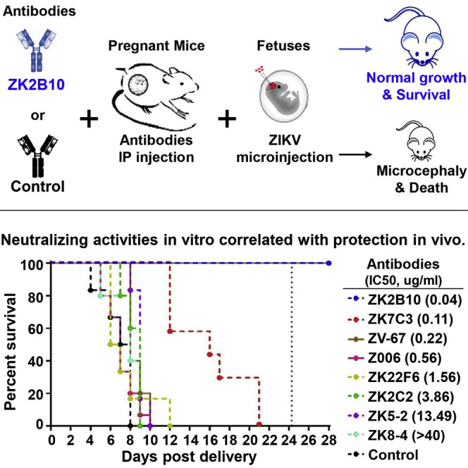
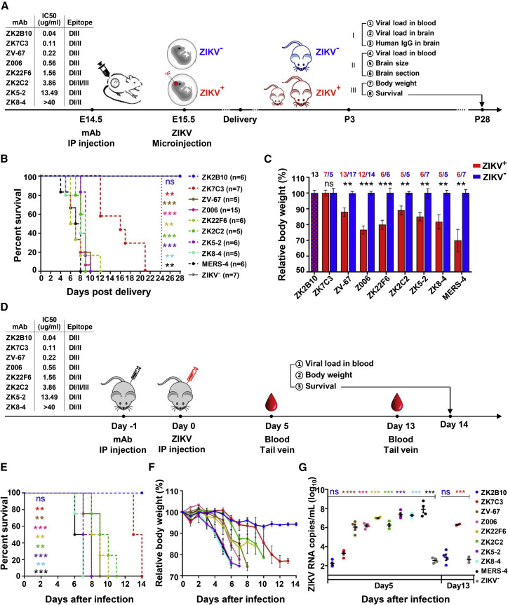
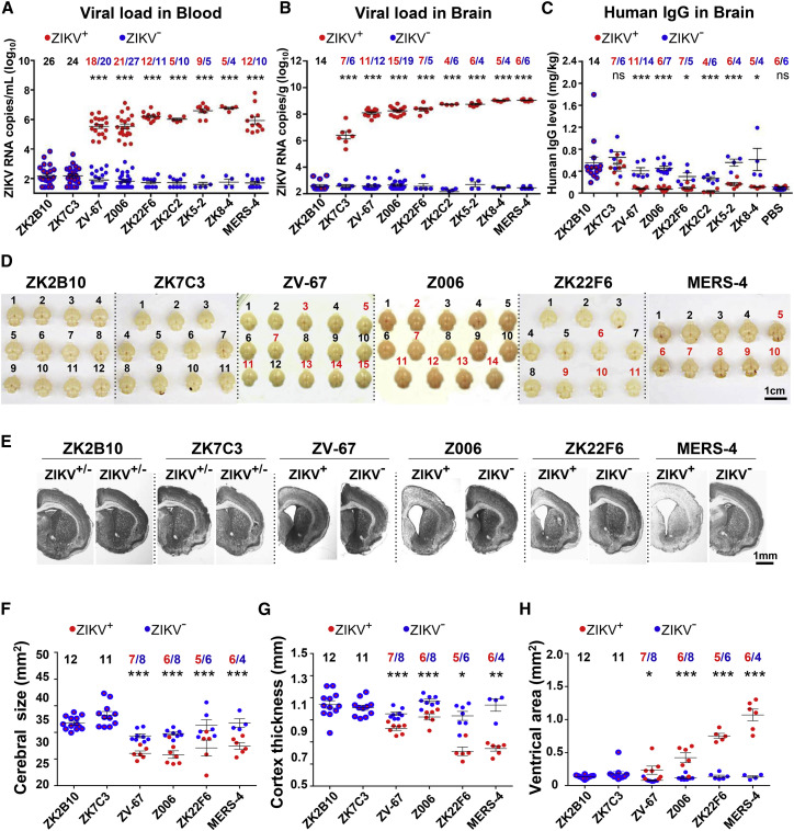
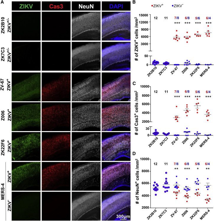
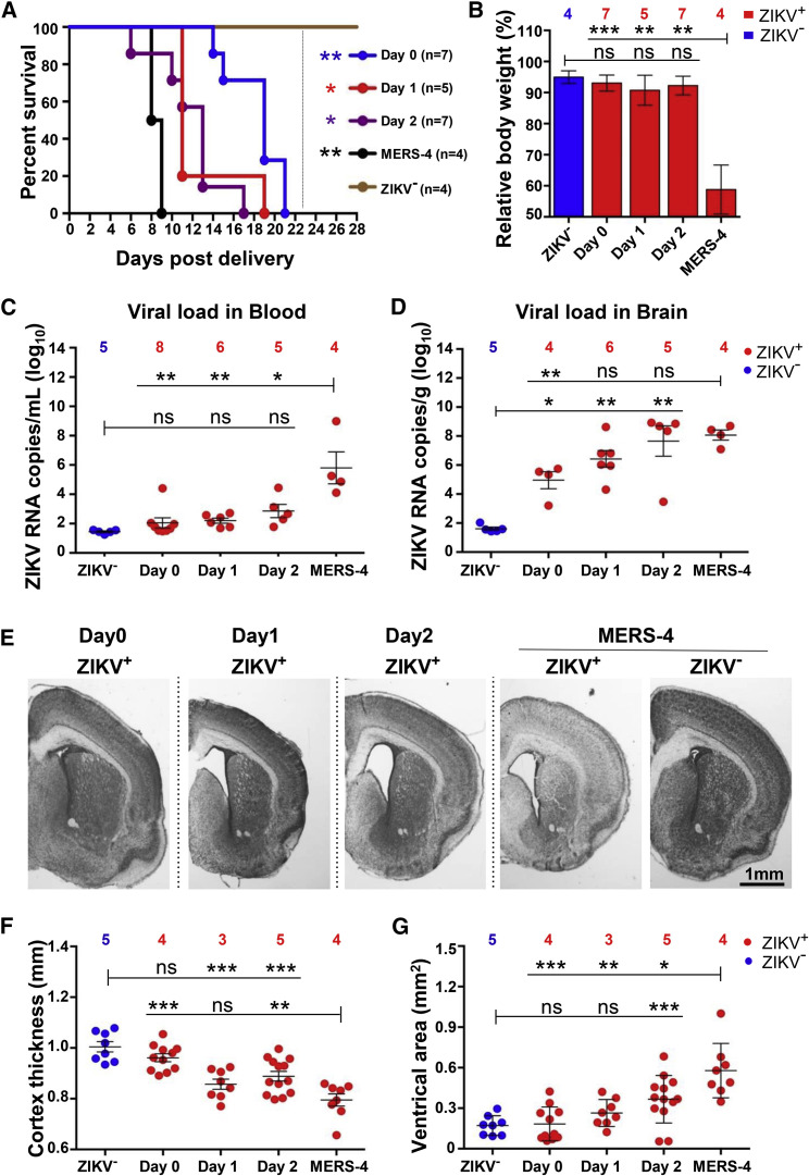
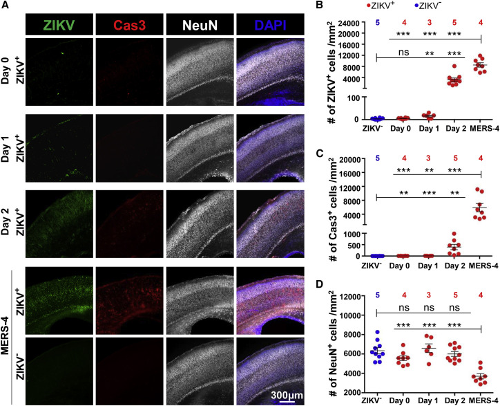
Zika virus (ZIKV) is a mosquito-transmitted flavivirus that is generally benign in humans. However, an emergent strain of ZIKV has become widespread, causing severe pre- and post-natal neurological defects. There is now an urgent need for prophylactic and therapeutic agents. To address this, we investigated six human monoclonal antibodies with ZIKV epitope specificity and neutralizing activity in mouse models of ZIKV infection and microcephaly. A single intraperitoneal injection of these antibodies conveyed distinct levels of adult and in utero protection from ZIKV infection, which closely mirrored their respective in vitro neutralizing activities. One antibody, ZK2B10, showed the most potent neutralization activity, completely protected uninfected mice, and markedly reduced tissue pathology in infected mice. Thus, ZK2B10 is a promising candidate for the development of antibody-based interventions and informs the rational design of ZIKV vaccine.

Keywords: Zika virus; epitope; microcephaly; neural progenitor cells; neutralizing antibody; protection; vaccine.


Conflict of interest statement

Patents have been filed for the isolated antibodies, and they are all available for collaboration in research and development through material transfer agreement.

Figures

None
Graphical abstract
Figure 1
Figure 1
Experimental Design to Evaluate the Prophylactic Potential of Six Human mAbs against ZIKV Infection in Developing Fetuses and AG6 Mice (A) Timeline for mAb injection, ZIKV inoculation, infant delivery, and the monitoring of a complementary set of clinical, virological, and neuropathological outcomes from embryonic day 14.5 to P28. The six mAbs tested are shown alongside their IC50 values and epitope specificities. ZIKV-inoculated fetuses and neonates are indicated by ZIKV+ in red; those left unexposed are indicated by ZIKV in blue. The cartoon mice on P3 include ZIKV+ (small and large indicated in red) and ZIKV (large indicated in blue); the size representations reflect potential body weight outcomes. Each mAb was tested, and the outcomes were monitored in three littermate neonates of three pregnant mice on P3 and P28. (B) Different levels of protection conferred by the six mAbs, shown with the number (n) of neonates monitored for survival in each mAb treatment group. (C) The body weight of neonates among the different mAb groups. ZIKV+ neonates indicated in red are presented as a percentage of the blue ZIKV littermates. The ZK2B10-treated group had no discernable differences between the ZIKV+ and ZIKV littermates and is, therefore, shown as a single red/blue checkered bar. The number of neonates for each analysis is indicated above the respective mAb, with ZIKV+ in red and ZIKV in blue. (D–G) Shown here: (D) timeline for mAb injection, ZIKV inoculation, and monitoring for (E) survival, (F) body weight, and (G) blood ZIKV RNA up to 14 days in AG6 mice. The body weight and ZIKV RNA in the whole blood derived from a single measurement showed distinct results among the study groups. The number of animals used in each group was four. All data are presented as mean ± SEM. p < 0.05; ∗∗p < 0.01; ∗∗∗p < 0.001; ns, no significant.
Figure 2
Figure 2
Marked Improvements in Virologic and Neuropathologic Outcomes in the Protected Mice (A and B) ZIKV RNA copies (A) in the blood and (B) in the brain of the neonatal mice at P3, as measured by qRT-PCR and presented as RNA copy equivalents per milliliter and per gram, respectively. (C) The levels of human IgG in the neonatal brains at P3 measured by ELISA and presented as milligram per kilogram brain. (D) Images of ZIKV+ and ZIKV littermate brains at P3. Red numbers represent ZIKV+ littermates in ZV-67-, Z006-, ZK22F6-, and MERS-4-treated groups. (E) Representative coronal sections of the brains with Nissl staining. Scale bar, 1 mm. (F–H) Quantitative analysis of (F) the cerebral size, (G) the cortex thickness, and (H) the ventricular area of the neonatal brains at P3 treated with indicated mAbs. Each red dot represents a ZIKV+; each blue dot represents a ZIKV neonate. The red dots with blue coating represent no discernable differences between ZIKV+ and ZIKV littermates in ZK2B10- and ZK7C3-treated groups. The numbers of neonates for each mAb analyses are indicated above each group, with ZIKV+ in red and ZIKV in blue. In (G) and (H), each dot represents the mean value of at least two slices from one neonate. All data are presented as mean ± SEM. p < 0.05; ∗∗p < 0.01; ∗∗∗p < 0.001; ns, no significant.
Figure 3
Figure 3
Brain Sections of Protected Animals Indicated Marked Reductions in Cell and Tissue Damage (A) Representative coronal section of cortices stained for ZIKV-infected cells (ZIKV antiserum indicated in green), apoptotic cells (Case3+ indicated in red), mature neurons (NeuN indicated in gray), and cellular DNA in the nuclei (DAPI indicated in blue). (B–D) Quantitative analyses for each cell type among the tested mAbs and an isotype control MERS-4. (B) Number of ZIKV+ cells per square millimeter. (C) Number of Cas3+ cells per square millimeter. (D) Number of NeuN+ cells per square millimeter. Each red dot represents a ZIKV+ neonate; each blue dot represents a ZIKV littermate. Red dots with blue coating represent no discernable differences between ZIKV+ and ZIKV littermates in ZK2B10- and ZK7C3-treated groups. Each dot represents the mean value of at least two slices from one neonate analyzed. The numbers of neonates for each analysis are indicated above each group, with ZIKV+ in red and ZIKV in blue. Scale bar, 300 μm. All data are presented as mean ± SEM. p < 0.05; ∗∗p < 0.01; ∗∗∗p < 0.001.
Figure 4
Figure 4
Evidence of Therapeutic Potential of ZK2B10 against ZIKV Infection in Developing Fetuses (A) Decreased survival when treatment administration is delayed from day 0 (E15.5) to day 1 (E16.5) or to day 2 (E17.5). The number (n) of neonates monitored for survival is indicated. (B–D) After treatment with ZK2B10 on day 0, day 1, or day 2, we analyzed ZIKV+ neonates (indicated in red) and ZIKV littermates (indicated in blue) at P3 for (B) body weight, (C) ZIKV RNA copies in the blood, and (D) ZIVK RNA copies in the brain. The number of neonates analyzed for each treatment time point is indicated above each group. (E–G) Shown here: (E) the representative coronal sections of the neonatal brains at P3 was visualized with Nissl staining and analyzed for (F) cortex thickness and (G) ventricular area. In (F) and (G), each dot represents the value of one slice. The total numbers of slices analyzed for ZIKV, day 0, day 1, day 2, and MERS-4 were 8, 11, 8, 13, and 8, respectively. The number of neonates analyzed for each treatment time point is indicated above each group. Scale bar, 1 mm. All data are presented as mean ± SEM. p < 0.05; ∗∗p < 0.01; ∗∗∗p < 0.001; ns, no significant.
Figure 5
Figure 5
ZK2B10 Reduced Brain Cell and Tissue Damage in Treated Mice (A) Following treatment with ZK2B10 on day 0, day 1, or day 2, representative coronal section of cortices stained for ZIKV-infected cells (ZIKV antiserum indicated in green), apoptotic cells (Cas3+ indicated in red), mature neurons (NeuN indicated in gray), and cellular DNA in the nuclei (DAPI indicated in blue). Scale bar, 300 μm. (B–D) Quantitative analysis for each cell type among different treatment points and an isotype control MERS-4. Each dot represents one slice. (B) Number of ZIKV+ cells per square millimeter. (C) Number of Cas3+ cells per square millimeter. (D) Number of NeuN+ cells per square millimeter. The total numbers of slices analyzed for ZIKV, day 0, day 1, day 2, and MERS-4 were 10, 8, 6, 10, and 8 in (B) and (D), and 10, 8, 6, 8, and 8 in (C), respectively. The total number of neonates analyzed is indicated above each group. All data are presented as mean ± SEM. ∗∗p < 0.01; ∗∗∗p < 0.001; ns, not significant.

Similar articles

Cited by

References

    1. Burton D.R., Hangartner L. Broadly Neutralizing Antibodies to HIV and Their Role in Vaccine Design. Annu. Rev. Immunol. 2016;34:635–659. - PMC - PubMed
    1. Corti D., Lanzavecchia A. Broadly neutralizing antiviral antibodies. Annu. Rev. Immunol. 2013;31:705–742. - PubMed
    1. Cugola F.R., Fernandes I.R., Russo F.B., Freitas B.C., Dias J.L., Guimarães K.P., Benazzato C., Almeida N., Pignatari G.C., Romero S., et al. The Brazilian Zika virus strain causes birth defects in experimental models. Nature. 2016;534:267–271. - PMC - PubMed
    1. Dang J., Tiwari S.K., Lichinchi G., Qin Y., Patil V.S., Eroshkin A.M., Rana T.M. Zika Virus Depletes Neural Progenitors in Human Cerebral Organoids through Activation of the Innate Immune Receptor TLR3. Cell Stem Cell. 2016;19:258–265. - PMC - PubMed
    1. Dejnirattisai W., Wongwiwat W., Supasa S., Zhang X., Dai X., Rouvinski A., Jumnainsong A., Edwards C., Quyen N.T.H., Duangchinda T., et al. A new class of highly potent, broadly neutralizing antibodies isolated from viremic patients infected with dengue virus. Nat. Immunol. 2015;16:170–177. - PMC - PubMed

Publication types

MeSH terms