. 2018 May;22(3):349-360.
doi: 10.4196/kjpp.2018.22.3.349. Epub 2018 Apr 25.

Anti-apoptotic effects of autophagy via ROS regulation in microtubule-targeted and PDGF-stimulated vascular smooth muscle cells

Affiliations

Anti-apoptotic effects of autophagy via ROS regulation in microtubule-targeted and PDGF-stimulated vascular smooth muscle cells

Hyun-Soo Park et al. Korean J Physiol Pharmacol. 2018 May.

Abstract

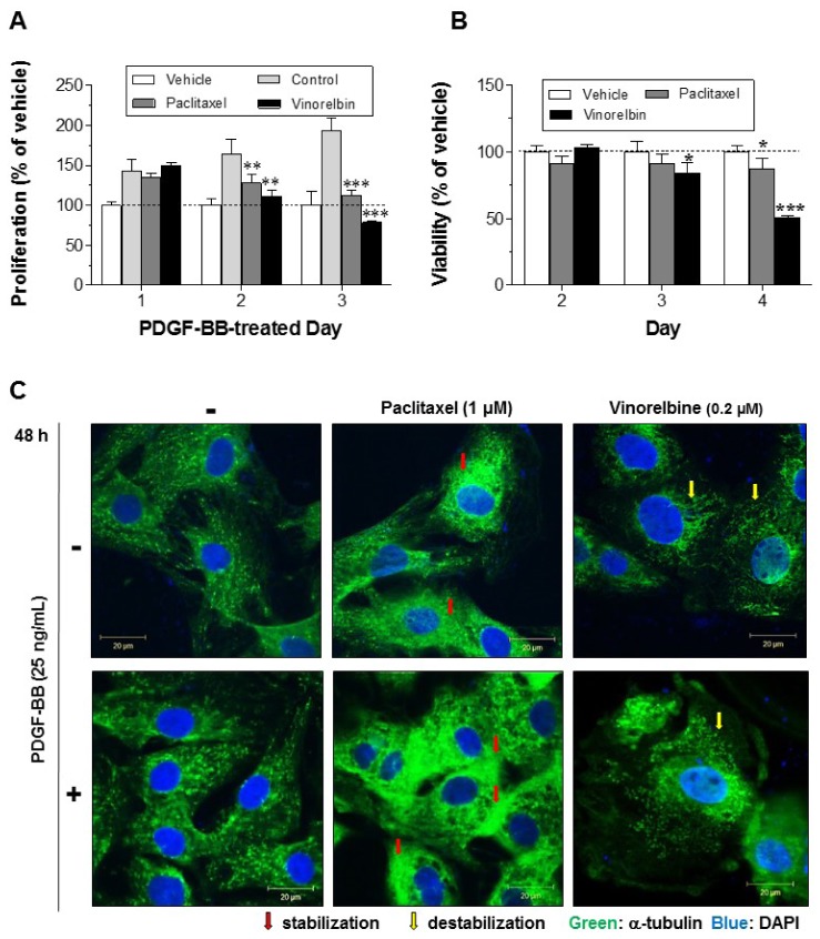
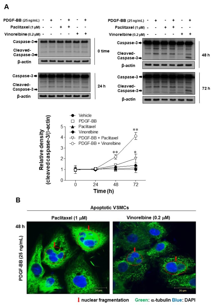
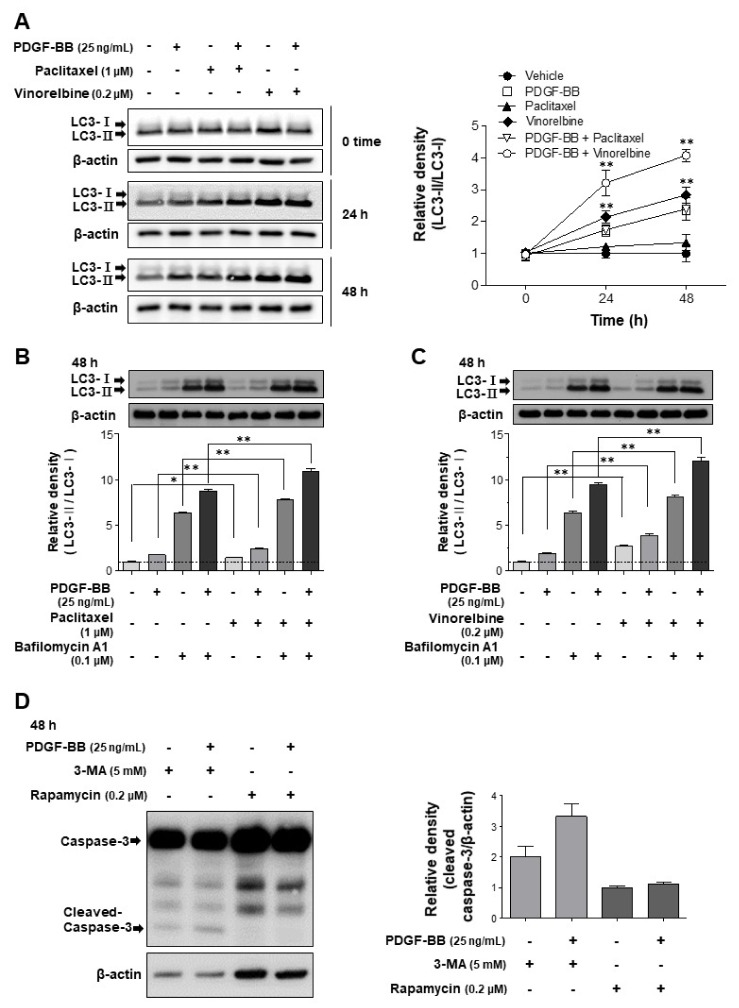
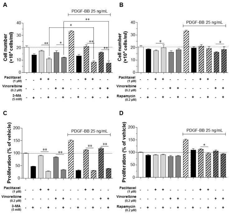
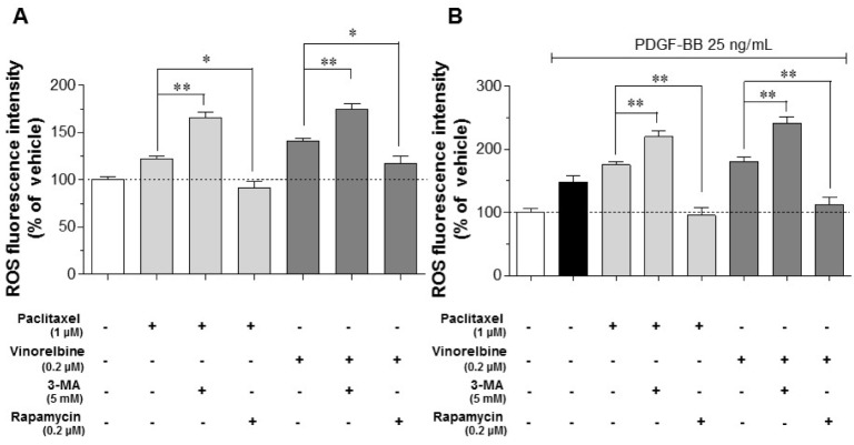
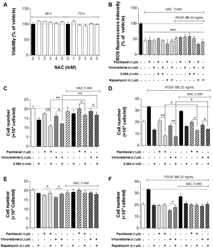
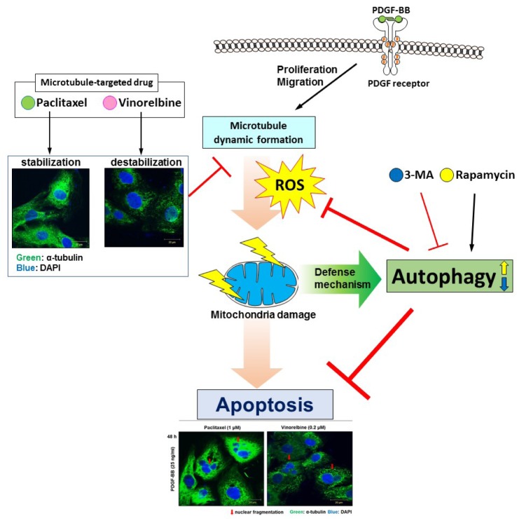
Autophagy has been studied as a therapeutic strategy for cardiovascular diseases. However, insufficient studies have been reported concerning the influence of vascular smooth muscle cells (VSMCs) through autophagy regulation. The aim of the present study was to determine the effects of VSMCs on the regulation of autophagy under in vitro conditions similar to vascular status of the equipped microtubule target agent-eluting stent and increased release of platelet-derived growth factor-BB (PDGF-BB). Cell viability and proliferation were measured using MTT and cell counting assays. Immunofluorescence using an anti-α-tubulin antibody was performed to determine microtubule dynamic formation. Cell apoptosis was measured by cleavage of caspase-3 using western blot analysis, and by nuclear fragmentation using a fluorescence assay. Autophagy activity was assessed by microtubule-associated protein light chain 3-II (LC-II) using western blot analysis. Levels of intracellular reactive oxygen species (ROS) were measured using H2DCFDA. The proliferation and viability of VSMCs were inhibited by microtubule regulation. Additionally, microtubule-regulated and PDGF-BB-stimulated VSMCs increased the cleavage of caspase-3 more than only the microtubule-regulated condition, similar to that of LC3-II, implying autophagy. Inhibitory autophagy of microtubule-regulated and PDGF-BB-stimulated VSMCs resulted in low viability. However, enhancement of autophagy maintained survival through the reduction of ROS. These results suggest that the apoptosis of conditioned VSMCs is decreased by the blocking generation of ROS via the promotion of autophagy, and proliferation is also inhibited. Thus, promoting autophagy as a therapeutic target for vascular restenosis and atherosclerosis may be a good strategy.

Keywords: Apoptosis; Autophagy; Proliferation; Reactive oxygen species; Vascular smooth muscle cell.


Conflict of interest statement

CONFLICTS OF INTEREST: The authors declare no conflicts of interest.

Figures

Fig. 1
Fig. 1. Effects of paclitaxel and vinorelbine on VSMC proliferation, viability and microtubule regulation.
Serum-starved VSMCs were incubated with 1 µM paclitaxel or 0.2 µM vinorelbine for 24 h followed by 25 ng/ml PDGF-BB-treatment for 24–2 h. VSMC proliferation and viability were evaluated using the MTT assay (A, B). Mean values of the vehicle group (0.1% DMSO) were set to 100%. Data are expressed as means±SEM (n = 3). *p<0.05, **p<0.01, ***p<0.001 vs. control (PDGF-BB alone) or vehicle. (C) Microtubules were observed by confocal microscopy. After cells were fixed with 4% formaldehyde and membrane-permeabilized using 0.25% Triton X-100, immunofluorescence staining was performed using anti-α-tubulin and anti-FITC antibodies. Nuclei were stained with DAPI. Red arrow indicates stabilization of microtubule and yellow arrow indicates destabilization of microtubule. Immunofluorescence images are representative of those obtained from three independent experiments (scale bar: 20 µm; nuclei: blue; α-tubulin: green).
Fig. 2
Fig. 2. Effects of microtubule regulation on caspase-3 in PDGF-BB-stimulated VSMCs.
Serum-starved VSMCs were incubated with 1 µM paclitaxel or 0.2 µM vinorelbine for 24 h followed by 25 ng/ml PDGF-BB treatment for 24–72 h. (A) VSMC apoptosis was evaluated according to the levels of caspase-3 cleavage (an apoptosis marker). The levels of full-length caspase 3 (35 kDa) and the cleaved fragment (17–19 kDa) were assessed by western blotting. The band densities were normalized to those of β-actin. The gel images shown are representative of those obtained from three independent experiments. The relative density was plotted by line graph. Mean values of the vehicle group (0.1% DMSO) were set to 1 fold. Data are expressed as means±SEM. *p<0.05, **p<0.01 vs. vehicle. (B) Immunofluorescence staining was performed using anti-α-tubulin and anti-FITC antibodies, and the VSMC nuclei were stained with DAPI (arrow: nuclear fragmentation; scale bar: 20 µm; nuclei: blue; α-tubulin: green).
Fig. 3
Fig. 3. Effects of autophagy via microtubule regulation and effects of caspase-3 cleavage via autophagy regulation in PDGF-BB-stimulated VSMCs.
Serum-starved VSMCs were incubated with 1 µM paclitaxel, 0.2 µM vinorelbine, 5 mM 3-MA (an autophagy inhibitor), or 0.2 µM rapamycin (an autophagy stimulator) for 24 h followed by 25 ng/ml PDGF-BB treatment for 0–48 h. Additionally, the cells were treated with 0.1 µM bafilomycin A1 for 4 h before the end of the reaction. Cell lysates applied to SDS-PAGE and subsequently immunoblotted. The band densities were normalized to those of β-actin. The gel images shown are representative of those obtained from three or five independent experiments. Data shown in graphs for relative density are expressed as means±SEM. (A) The conversion of LC3-I to LC3-II was measured as an autophagy key marker in microtubule-regulated VSMCs before and after (24–48 h) PDGF-BB treatment. Mean values of the vehicle group (0.1% DMSO) were set to 1 fold. **p<0.01 vs. vehicle. (B, C) Measurement of autophagic flux. Bafilomycin A1 (0.1 µM) was added to the vehicle (0.1% DMSO)-treated, microtubule-regulated (paclitaxel or vinorelbine), or PDGF-BB-stimulated VSMCs 4 h prior to harvest, and accumulation of LC3-II was measured by western blotting. The mean value of vehicle was set to 1 fold. *p<0.05, **p<0.01 vs. the indicated group. (D) The changes of apoptosis by autophagy regulation were tested by the measurement of caspase-3 cleavage levels. Mean values of the only rapamycin-treated group were set to 1 fold.
Fig. 4
Fig. 4. Effects of the regulation of microtubules and autophagy on the proliferation and viability in PDGF-BB-stimulated VSMCs.
Serum-starved VSMCs were incubated with 1 µM paclitaxel, 0.2 µM vinorelbine, 5 mM 3-MA (autophagy inhibitor), or 0.2 µM rapamycin (autophagy stimulator) for 24 h followed by 25 ng/ml PDGF-BB treatment for 48 h. Microtubule-regulated and PDGF-BB-stimulated VSMCs viability and proliferation by autophagy regulation were determined using the cell counting assay (A, B) and MTT assay (C, D). Data are expressed as means±SEM (n=3). *p<0.05, **p<0.01 vs. the indicated group.
Fig. 5
Fig. 5. Intracellular ROS levels through microtubule regulation and autophagy in PDGF-BB-stimulated VSMCs.
Serum-deprived VSMCs were incubated with 1 µM paclitaxel, 0.2 µM vinorelbine, 5 mM 3-MA (autophagy inhibitor), or 0.2 µM rapamycin (autophagy stimulator) for 24 h followed by 25 ng/ml PDGF-BB treatment for 48 h. After stimulation, the cells were stained with 20 µM H2DCFDA for 30 min at 37℃, and the fluorescence intensities were measured. (A) ROS levels of microtubule- and autophagy-regulated VSMCs. (B) ROS levels of microtubule- and autophagy-regulated, and PDGF-BB-stimulated VSMCs. Mean values of the vehicle group (0.1% DMSO) were set to 100%. Data are expressed as means±SEM (n=3). *p<0.05, **p<0.01 vs. the indicated group.
Fig. 6
Fig. 6. Effects of the regulation of intracellular ROS, microtubule and autophagy on proliferation and viability in PDGF-BB-stimulated VSMCs.
Serum-deprived VSMCs were incubated with 1 µM paclitaxel, 0.2 µM vinorelbine, 5 mM 3-MA (autophagy inhibitor), 0.2 µM rapamycin (autophagy stimulator), or 5 mM NAC (ROS scavenger) for 24 h followed by 25 ng/ml PDGF-BB treatment for 48 h. (A) Effects of NAC on cell viability. VSMCs cultured in serum-free medium were incubated with 1–5 mM NAC for the indicated periods, followed by subjecting the cells to the MTT assay. (B) Effects of NAC (5 mM) on ROS reduction under the regulation of microtubule and autophagy. The conditioned VSMCs were cultured with 5 mM NAC, and the ROS levels were measured using the H2DCFDA assay. Mean values of the vehicle group (0.1% DMSO) were set to 100%. ***p<0.001 vs. vehicle. The proliferation and viability of microtubule-regulated, autophagy-inhibited (C, D), and autophagy-stimulated (E, F), with or without PDGF-BB-treated VSMCs under the regulation of ROS were determined using the cell counting assay. Data are expressed as means±SEM (n=3). *p<0.05, **p<0.01 vs. the indicated group.
Fig. 7
Fig. 7. Proposed scheme for the antiapoptotic effect of autophagy via ROS regulation in microtubule-regulated and PDGF-BB-stimulated VSMCs.
PDGF receptor activation induces microtubule dynamic formation in VSMCs for cell proliferation and migration, which can be inhibited by microtubuletargeted drugs such as paclitaxel and vinorelbine, resulting in excessive production of ROS. This causes mitochondria damage, followed by apoptosis. Rapamycin as an autophagy activator can prevent excessive ROS production and apoptosis, resulting in cell viable. Thus, autophagy can act as a defense mechanism in microtubule-regulated VSMCs, and a regulator of apoptosis of conditioned VSMCs.

References

    1. Chistiakov DA, Orekhov AN, Bobryshev YV. Vascular smooth muscle cell in atherosclerosis. Acta Physiol (Oxf) 2015;214:33–50. - PubMed
    1. Shioi A, Ikari Y. Plaque calcification during atherosclerosis progression and regression. J Atheroscler Thromb. 2018;25:294–303. - PMC - PubMed
    1. van der Wal AC, Becker AE. Atherosclerotic plaque rupture— pathologic basis of plaque stability and instability. Cardiovasc Res. 1999;41:334–344. - PubMed
    1. Ramji DP, Davies TS. Cytokines in atherosclerosis: key players in all stages of disease and promising therapeutic targets. Cytokine Growth Factor Rev. 2015;26:673–685. - PMC - PubMed
    1. Ross R. Growth factors in the pathogenesis of atherosclerosis. Acta Med Scand Suppl. 1987;715:33–38. - PubMed