. 2018 Apr 17:12:147.
doi: 10.3389/fnhum.2018.00147. eCollection 2018.

Descending Inputs to Spinal Circuits Facilitating and Inhibiting Human Wrist Flexors

Affiliations

Descending Inputs to Spinal Circuits Facilitating and Inhibiting Human Wrist Flexors

Stefane A Aguiar et al. Front Hum Neurosci. .

Abstract

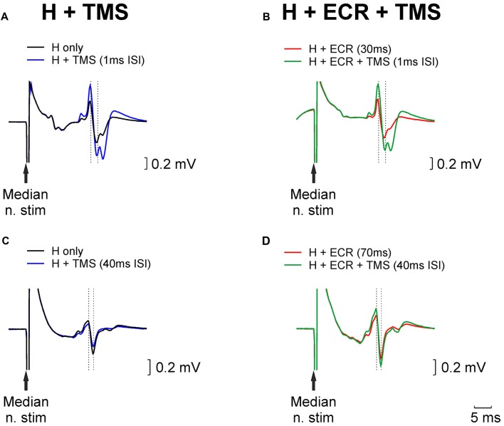
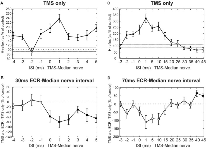
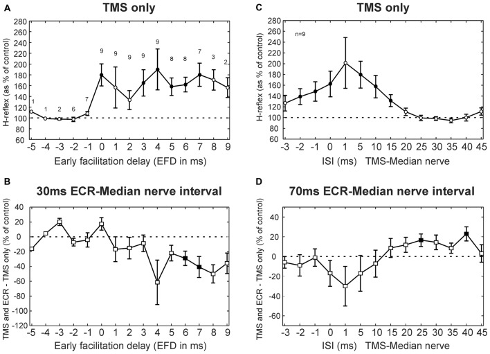
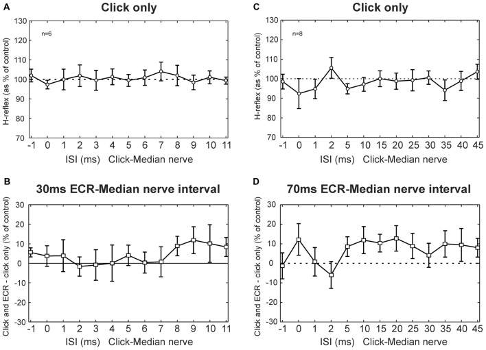
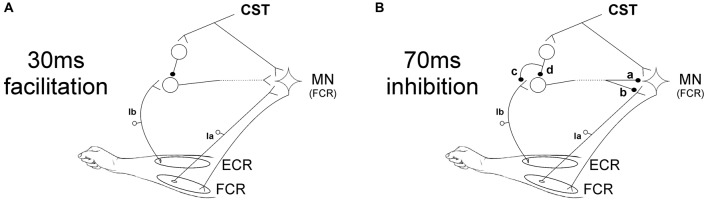
Recently we reported in humans that electrical stimulation of the wrist extensor muscle extensor carpi radialis (ECR) could facilitate or suppress the H reflex elicited in flexor carpi radialis (FCR), for inter-stimulus intervals (ISIs) of 30 ms or 70 ms, respectively. The facilitation at 30 ms may be produced by both flexor afferents and extensor Ib afferents acting on a spinal circuit; the origin of the suppression at 70 ms is less certain. In this study, we investigated possible descending inputs to these systems. We used magnetic stimulation of the contralateral primary motor cortex, and click sound stimulation, to activate the corticospinal and the reticulospinal tracts respectively, and measured the effects on the H reflex conditioned by ECR stimulation. Corticospinal inputs reduced both the 30 ms facilitation and 70 ms suppression, indicating corticospinal inhibition of both circuits. By contrast, we failed to show any effect of clicks, either on the H reflex or on its modulation by ECR stimulation. This suggests that click-activated reticulospinal inputs to these circuits may be weak or absent.

Keywords: H-reflex; Ib afferents; click sound stimulation; corticospinal tract; reticulospinal tract; transcranial magnetic stimulation.


Figures

Figure 1
Figure 1
Examples of H reflex conditioning. (A) Black trace illustrates an unconditioned H reflex. Blue trace shows the H reflex conditioned by TMS delivered 1 ms before the median nerve stimulus; a clear facilitation is evident. (B) Red trace shows the H reflex conditioned by extensor carpi radialis (ECR) stimulation 30 ms before the median nerve stimulus. This facilitated the reflex; the median nerve intensity was reduced until the reflex amplitude approximately matched the unconditioned reflex (black trace, Panel A). Green trace illustrates the effect of conditioning this reflex with TMS (same timing relative to median nerve as in Panel A). (C,D) As for (A,B), but now illustrating ECR stimulation delivered 70 ms before the median nerve stimulus, which produced a reflex suppression, necessitating an increase in stimulus intensity to match the reflex amplitude. TMS in (C,D), was delivered 40 ms before the median nerve stimulus.
Figure 2
Figure 2
Transcranial magnetic stimulation (TMS) results from single subjects. (A) Effects of TMS on the flexor carpi radialis (FCR) H-reflex at different inter-stimulus intervals (ISIs) from a single subject. (B) Difference between the effect of TMS on the H reflex, and the effect of TMS on the H reflex conditioned by ECR stimulation 30 ms before the median nerve shock. Same subject as (A). (C) Effects of TMS on the FCR H-reflex, for different ISIs and a different subject from (A). (D) Difference between effect of TMS on H reflex, and on H reflex conditioned by ECR stimulation 70 ms before the median nerve shock. Same subject as (D). In (A,C), the peak-peak amplitude of the H reflex is plotted as a function of ISI, as a percentage of the size of the unconditioned H reflex. Filled symbols show points significantly different from 100% (A,C) or 0% (B,D). Error bars indicate standard error of the mean.
Figure 3
Figure 3
TMS results averaged across subjects. (A) Effects of TMS on the FCR H-reflex at different early facilitation delays (EFD). Numbers above each result display the number of subjects contributing to each data point. (B) Difference between the effect of TMS on the H reflex, and the effect of TMS on the H reflex conditioned by ECR stimulation 30 ms before the median nerve shock. Numbers of subjects contributing at each interval are as in (A). (C) Effects of TMS on the FCR H-reflex at different ISIs. (D) Difference between effect of TMS on H reflex, and on H reflex conditioned by ECR stimulation 70 ms before the median nerve shock. Filled symbols show responses significantly different from 100% (A,C) or 0% (B,D). Error bars indicate standard error of the mean. (C,D) are both averaged over n = 9 subjects.
Figure 4
Figure 4
Clicks results averaged across subjects. (A) Effects of clicks on the FCR H-reflex at different ISIs. (B) Between the effect of click on the H reflex, and the effect of click on the H reflex conditioned by ECR stimulation 30 ms before the median nerve shock which elicited the H reflex. (A,B) are averaged over n = 6 subjects. (C) Effects of clicks on the FCR H-reflex at different ISIs (note different time scale from A). (D) Difference between effect of click on H reflex, and on H reflex conditioned by ECR stimulation 70 ms before the median nerve shock. (C,D) are averaged over n = 8 subjects. In no cases in any plot were responses significantly different from control. Error bars indicate standard error of the mean.
Figure 5
Figure 5
Schematic representation of spinal circuits. (A) Corticospinal tract (CST) input to spinal circuit generating facilitation of the FCR H-reflex at 30 ms ECR-Median nerve interval. The CST inhibits Ib interneurons which excite FCR motorneurons (MNs). (B) CST input to spinal circuit generating suppression of the FCR H-reflex at 70 ms ECR-Median nerve interval. The components of this circuit are uncertain, and several possibilities are shown. The interneuron excited by Ib afferents may inhibit the FCR motorneurons post-synaptically (a) or pre-synaptically (b). The CST may inhibit this interneuron either via pre-synaptic inhibition to its Ib inputs (c) or via post-synaptic inhibition of the interneuron (d).

Similar articles

Cited by

References

    1. Aguiar S. A., Baker S. N. (2018). Convergent spinal circuits facilitating human wrist flexors. J. Neurosci. [Epub ahead of print]. 10.1523/JNEUROSCI.1870-17.2018 - DOI - PMC - PubMed
    1. Baker S. N. (2011). The primate reticulospinal tract, hand function and functional recovery. J. Physiol. 589, 5603–5612. 10.1113/jphysiol.2011.215160 - DOI - PMC - PubMed
    1. Baker S. N., Zaaimi B., Fisher K. M., Edgley S. A., Soteropoulos D. S. (2015). Pathways mediating functional recovery. Prog. Brain Res. 218, 389–412. 10.1016/bs.pbr.2014.12.010 - DOI - PubMed
    1. Baldissera F., Cavallari P. (1993). Short-latency subliminal effects of transcranial magnetic stimulation on forearm motoneurones. Exp. Brain Res. 96, 513–518. 10.1007/bf00234119 - DOI - PubMed
    1. Berardelli A., Day B. L., Marsden C. D., Rothwell J. C. (1987). Evidence favouring presynaptic inhibition between antagonist muscle afferents in the human forearm. J. Physiol. 391, 71–83. 10.1113/jphysiol.1987.sp016726 - DOI - PMC - PubMed

LinkOut - more resources