. 2018 Apr 6;9(26):18002-18017.
doi: 10.18632/oncotarget.24649.

Cold atmospheric plasma as a potential tool for multiple myeloma treatment

Affiliations

Cold atmospheric plasma as a potential tool for multiple myeloma treatment

Dehui Xu et al. Oncotarget. .

Abstract

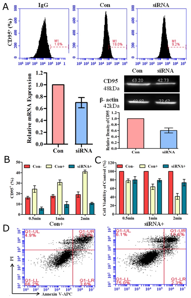
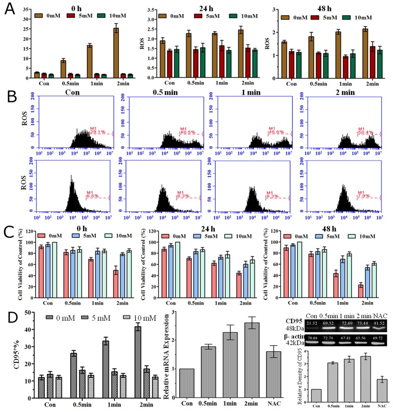
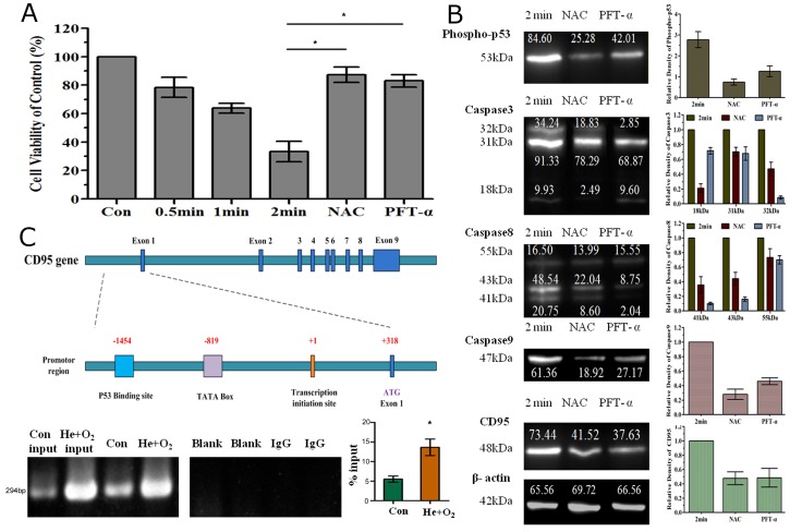
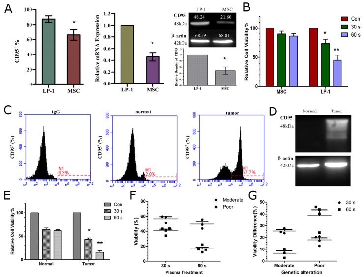
Multiple myeloma (MM) is a fatal and incurable hematological malignancy thus new therapy need to be developed. Cold atmospheric plasma, a new technology that could generate various active species, could efficiently induce various tumor cells apoptosis. More details about the interaction of plasma and tumor cells need to be addressed before the application of gas plasma in clinical cancer treatment. In this study, we demonstrate that He+O2 plasma could efficiently induce myeloma cell apoptosis through the activation of CD95 and downstream caspase cascades. Extracellular and intracellular reactive oxygen species (ROS) accumulation is essential for CD95-mediated cell apoptosis in response to plasma treatment. Furthermore, p53 is shown to be a key transcription factor in activating CD95 and caspase cascades. More importantly, we demonstrate that CD95 expression is higher in tumor cells than in normal cells in both MM cell lines and MM clinical samples, which suggests that CD95 could be a favorable target for plasma treatment as it could selectively inactivate myeloma tumor cells. Our results illustrate the molecular details of plasma induced myeloma cell apoptosis and it shows that gas plasma could be a potential tool for myeloma therapy in the future.

Keywords: CD95; cold atmospheric plasma; multiple myeloma; reactive oxygen species; selective inactivation.


Conflict of interest statement

CONFLICTS OF INTEREST The authors declare no conflicts of interest.

Figures

Figure 1
Figure 1. Characteristics of the plasma generation system
(A) Schematic representation of the plasma jet. (B) Photographs of He plasma and He+O2 plasmas. (C) Monitoring of the applied voltage and current during He+O2 plasma generation. (D) Emission spectra of different He+O2 plasmas detected by the spectrometer. Some unique spectral lines (OH, N2, He and O) in the He+0.5% O2 plasmas are marked. (E) The intensity of several characteristic spectral lines are shown with varying O2 percentage in the working gas.
Figure 2
Figure 2. Cell viability and apoptosis after plasma treatment for different durations
(A) Analysis of cell viability 24 h and 48 h after He plasma treatment for different durations. (B) Analysis of cell viability 24 h after He+O2 plasma for different durations. (C) Cell apoptosis analyzed by flow cytometry after He+0.5%O2 plasma treatment for 0.5, 1, and 2 min.
Figure 3
Figure 3. MMP, lysosomal leakage and caspase activation induced by plasma treatment
(A, B) LP-1 cells stained with a reporter dye, JC-1, for mitochondrial condition are detected by (A) fluorescence microscopy and (B) flow cytometry. (C) Analysis of lysosomal leakage in LP-1 cells indicated by fluorescence staining with Lucifer yellow after plasma treatment for 0.5, 1 and 2 min. (D) The activity of caspase3/8/9 was measured with a Caspase Colorimetric Assay Kit 3 h and 6 h after plasma treatment.
Figure 4
Figure 4. Analysis of apoptosis-related protein array and CD95 expression after plasma treatment
(A) Cell apoptotic apoptosis-related protein array detection was performed 24 h after He+O2 plasma treatment for 1 min. (B) CD95 expression was measured by flow cytometry 24 h after He+O2 plasma treatment for 0.5 min, 1 min and 2 min. IgG was used as the isotype control of CD95. Percentage of CD95 expression and MFI ratio were expressed in M±SD. * indicates p<0.05, ** indicates p<0.01
Figure 5
Figure 5. Down-regulation of CD95 expression by siRNA reduced plasma-induced cell apoptosis
(A) CD95 expression detected by flow cytometry, real-time PCR and western blotting after siRNA-mediated knockdown for 48 h. IgG is the isotype control. Con indicates transfection of control scramble siRNA compared to CD95-targeting siRNA. (B, C) CD95 expression (B) and corresponding cell viability (C) 24 h after plasma treatment for 0.5 min, 1 min and 2 min. Con- and Con+ groups were transfected with control scramble siRNA and treated without or with He+O2 plasma. The siRNA+ group represents cells treated with He+O2 plasma after knockdown of CD95 by siRNA. (D) Cell apoptosis was detected 24 h after He+O2 plasma treatment for 1 min following transfection. * indicates p<0.05.
Figure 6
Figure 6. Extracellular and intracellular ROS, cell viability and CD95 expression were measured after He+O2 plasma treatment with ROS scavenger (NAC)
(A) Extracellular ROS level detected with a microplate reader at 0 h, 24 h and 48 h after plasma treatment for 0.5, 1 and 2 min with or without NAC. (B) Intracellular ROS level detected with a flow cytometer 24 h after plasma treatment without (above) or with (below) NAC. (C) Cell viability measured by CellTiter-Glo assay at 0 h, 24 h and 48 h after plasma treatment for 0.5, 1 and 2 min with or without NAC. (D) CD95 expression was detected by flow cytometry (left) and real-time PCR (middle) 24 h after He+O2 treatment and by western blot (right) 48 h after plasma treatment with or without NAC.
Figure 7
Figure 7. Involvement of p53 in CD95-mediated cell apoptosis by He+O2 plasma treatment
(A) Analysis of cell viability after plasma treatment for 0.5, 1 and 2 min with or without NAC or PFT-α (p53 inhibitor). (B) Western blot analysis of phospho-p53, caspase3/8/9 and CD95 expression after plasma treatment. NAC and PFT-α indicates that the ROS scavenger and p53 inhibitor were added respectively before plasma treatment for 2 min. (C) Interaction of p53 and CD95 promoter region in response to plasma treatment. The top panel shows the illustration of the CD95 promoter region and p53 binding site. The bottom panel shows the results of ChIP assay for p53 and CD95 promoter detected by RT-PCR and real-time PCR. * indicates p<0.05.
Figure 8
Figure 8. Differential CD95 expression and sensitivity to plasma of cell lines and patient samples
(A) CD95 expression in MM tumor cells and normal cells (MSC) detected by flow cytometry, real-time PCR and western blotting. (B) Sensitivity of tumor and normal cells to plasma treatment assessed by cell viability assay. (C, D) CD95 expression detected by flow cytometry (C) and western blotting (D) in a representative patient sample. (E) Sensitivity of tumor and normal cells derived from patients assessed by cell viability assay 24 h after plasma treatment. (F) Sensitivity of MM tumor cells derived from patients with moderate and poor prognosis (analyzed by FISH) in response to plasma treatment, (G) Difference in viability of tumor and normal cells after plasma treatment in samples from patients with moderate and poor prognoses. * indicates p<0.05, ** indicates p<0.01.

References

    1. Podar K, Chauhan D, Anderson KC. Bone marrow microenvironment and the identification of new targets for myeloma therapy. Leukemia. 2009;23:10–24. - PMC - PubMed
    1. Ludwig H, Miguel J, Dimopoulos M, Palumbo A, Sanz RG, Powles R, Lentzsch S, Chen WM, Hou J, Jurczyszyn A. International myeloma working group recommendations for global myeloma care. Leukemia. 2014;28:981–992. - PubMed
    1. Ocio EM, Richardson PG, Rajkumar SV, Palumbo A, Mateos MV, Orlowski R, Kumar S, Usmani S, Roodman D, Niesvizky R. New drugs and novel mechanisms of action in multiple myeloma in 2013: A report from the International Myeloma Working Group (IMWG) Leukemia. 2014;28:525–542. - PMC - PubMed
    1. Laubach J, Garderet L, Mahindra A, Gahrton G, Caers J, Sezer O, Voorhees P, Leleu X, Johnsen H, Streetly M. Management of relapsed multiple myeloma: recommendations of the International Myeloma Working Group. Leukemia. 2016;30:1005–1017. - PubMed
    1. Trachootham D, Alexandre J, Huang P. Targeting cancer cells by ROS-mediated mechanisms: a radical therapeutic approach? Nature reviews Drug discovery. 2009;8:579–591. - PubMed