Intraocular Lens Calcification: Clinico-pathological Report of Two Cases and Literature Review
- PMID: 29719650
- PMCID: PMC5905315
- DOI: 10.4103/jovr.jovr_36_16
Intraocular Lens Calcification: Clinico-pathological Report of Two Cases and Literature Review
Abstract
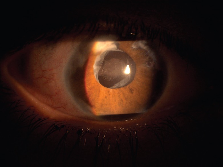
Purpose: We report the clinicopathological features and surgical outcomes of two cases of intraocular lens (IOL) calcification along with a review of the current literature.
Case report: The first patient was a 53-year-old woman with diabetes mellitus (type 2) who underwent phacoemulsification with posterior chamber IOL insertion (PCIOL), and pars plana vitrectomy. Significant clouding of the IOL was first noted after 1.5 years, and the IOL was replaced with an Artisan lens. The second patient was a 22-year-old woman with Vogt-Koyanagi-Harada syndrome; she underwent a lensectomy, PCIOL, and pars plana vitrectomy. IOL opacification was first noted 4.5 years after the initial surgery and the IOL was extracted.
Conclusion: The calcification of each IOL was confirmed by a pathologist. Further studies are required to determine the primary causes and mechanisms of the calcification of biomaterials including IOLs.
Keywords: Calcification; Intraocular Lens; Pathology.
Conflict of interest statement
There are no conflicts of interest.
Figures


Similar articles
-
Akreos Adapt AO Intraocular lens opacification after vitrectomy in a diabetic patient: a case report and review of the literature.BMC Ophthalmol. 2016 Jun 8;16:82. doi: 10.1186/s12886-016-0268-3. BMC Ophthalmol. 2016. PMID: 27277708 Free PMC article. Review.
-
Pars plana vitrectomy for posteriorly dislocated intraocular lenses: risk factors and surgical approach.Int Ophthalmol. 2021 Jan;41(1):221-229. doi: 10.1007/s10792-020-01570-7. Epub 2020 Sep 11. Int Ophthalmol. 2021. PMID: 32915391
-
Combined pars plana lensectomy-vitrectomy with open-loop flexible anterior chamber intraocular lens (AC IOL) implantation for subluxated lenses.Trans Am Ophthalmol Soc. 2000;98:247-51; discussion 251-3. Trans Am Ophthalmol Soc. 2000. PMID: 11190027 Free PMC article.
-
Unsutured posterior chamber lens implantation in eyes requiring lens extraction at the time of pars plana vitrectomy with silicone oil tamponade.J Cataract Refract Surg. 2004 Jan;30(1):161-7. doi: 10.1016/S0886-3350(03)00650-3. J Cataract Refract Surg. 2004. PMID: 14967285
-
Systematic review of potential causes of intraocular lens opacification.Clin Exp Ophthalmol. 2020 Jan;48(1):89-97. doi: 10.1111/ceo.13650. Epub 2019 Oct 22. Clin Exp Ophthalmol. 2020. PMID: 31581356
Cited by
-
Physicochemical Analysis of Sediments Formed on the Surface of Hydrophilic Intraocular Lens after Descemet's Stripping Endothelial Keratoplasty.Materials (Basel). 2020 Sep 17;13(18):4145. doi: 10.3390/ma13184145. Materials (Basel). 2020. PMID: 32957729 Free PMC article.
-
A rare intraocular lens surface foreign body during phacoemulsification surgery: A case report.Medicine (Baltimore). 2021 Jan 22;100(3):e24391. doi: 10.1097/MD.0000000000024391. Medicine (Baltimore). 2021. PMID: 33546080 Free PMC article.
-
Clinical Characteristics of Patients with Intraocular Lens Calcification after Pars Plana Vitrectomy.Diagnostics (Basel). 2023 Jun 1;13(11):1943. doi: 10.3390/diagnostics13111943. Diagnostics (Basel). 2023. PMID: 37296795 Free PMC article.
-
Corneal edema associated with degenerating Soemmering ring cataract: Clinical-pathologic correlation.Am J Ophthalmol Case Rep. 2022 Nov 11;28:101738. doi: 10.1016/j.ajoc.2022.101738. eCollection 2022 Dec. Am J Ophthalmol Case Rep. 2022. PMID: 36393913 Free PMC article.
References
-
- Apple DJ, Mamalis N, Olson RJ, Kincaid MC. Baltimore: Williams & Wilkins; 1989. IOLs: Evolution, designs, complications, and pathology.
-
- Mamalis N. Hydrophilic acrylic intraocular lenses. J Cataract Refract Surg. 2001;27:1339–1340. - PubMed
-
- Dorey MW, Brownstein S, Hill VE, Mathew B, Botton G, Kertes PJ, et al. Proposed pathogenesis for the delayed postoperative opacification of the hydroview hydrogel intraocular lens. Am J Ophthalmol. 2003;135:591–598. - PubMed
-
- Werner L, Apple DJ, Escobar-Gomez M. Postoperative deposition of calcium on the surface of a hydrogel intraocular lens. Ophthalmology. 2000;107:2179–2185. - PubMed
-
- Izak AM, Werner L, Pandey SK, Apple DJ. Calcification of modern foldable hydrogel intraocular lens designs. Eye. 2003;17:393–406. - PubMed
Publication types
LinkOut - more resources
Full Text Sources
Other Literature Sources
Miscellaneous