. 2018 May 2;18(1):507.
doi: 10.1186/s12885-018-4387-5.

C-Cbl reverses HER2-mediated tamoxifen resistance in human breast cancer cells

Affiliations

C-Cbl reverses HER2-mediated tamoxifen resistance in human breast cancer cells

Wei Li et al. BMC Cancer. .

Abstract

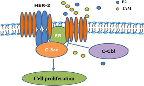
Background: Tamoxifen is a frontline therapy for estrogen receptor (ER)-positive breast cancer in premenopausal women. However, many patients develop resistance to tamoxifen, and the mechanism underlying tamoxifen resistance is not well understood. Here we examined whether ER-c-Src-HER2 complex formation is involved in tamoxifen resistance.

Methods: MTT and colony formation assays were used to measure cell viability and proliferation. Western blot was used to detect protein expression and protein complex formations were detected by immunoprecipitation and immunofluorescence. SiRNA was used to examine the function of HER2 in of BT474 cells. An in vivo xenograft animal model was established to examine the role of c-Cbl in tumor growth.

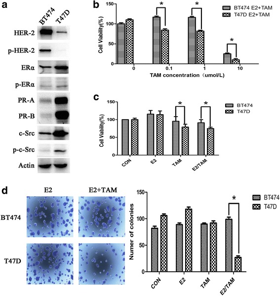
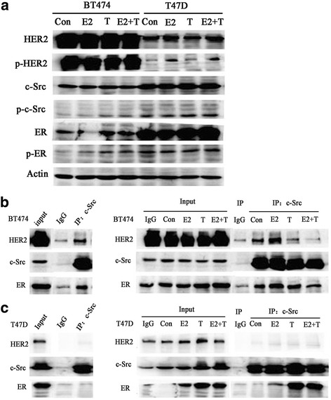
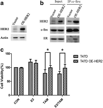
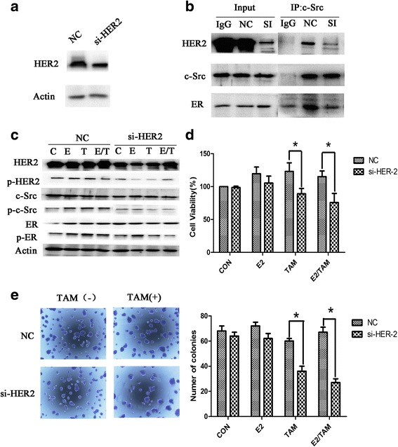
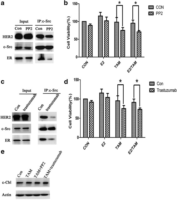
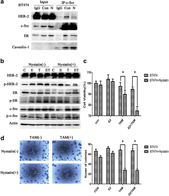
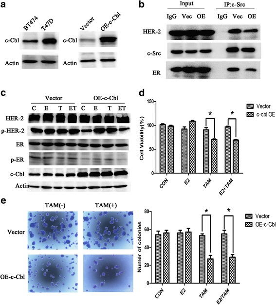
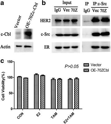
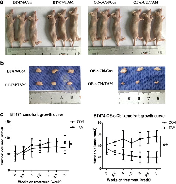
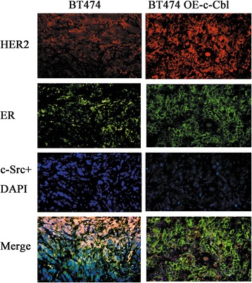
Results: MTT and colony formation assay showed that BT474 cells are resistant to tamoxifen and T47D cells are sensitive to tamoxifen. Immunoprecipitation experiments revealed ER-c-Src-HER2 complex formation in BT474 cells but not in T47D cells. However, ER-c-Src-HER2 complex formation was detected after overexpressing HER2 in T47D cells and these cells were more resistant to tamoxifen. HER2 knockdown by siRNA in BT474 cells reduced ER-c-Src-HER2 complex formation and reversed tamoxifen resistance. ER-c-Src-HER2 complex formation was also disrupted and tamoxifen resistance was reversed in BT474 cells by the c-Src inhibitor PP2 and HER2 antibody trastuzumab. Nystatin, a lipid raft inhibitor, reduced ER-c-Src-HER2 complex formation and partially reversed tamoxifen resistance. ER-c-Src-HER2 complex formation was disrupted by overexpression of c-Cbl but not by the c-Cbl ubiquitin ligase mutant. In addition, c-Cbl could reverse tamoxifen resistance in BT474 cells, but the ubiquitin ligase mutant had no effect. The effect of c-Cbl was validated in BT474 tumor-bearing nude mice in vivo. Immunofluorescence also revealed ER-c-Src-HER2 complex formation was reduced in tumor tissues of nude mice with c-Cbl overexpression.

Conclusions: Our results suggested that c-Cbl can reverse tamoxifen resistance in HER2-overexpressing breast cancer cells by inhibiting the formation of the ER-c-Src-HER2 complex.

Keywords: Breast cancer; C-Cbl; HER2; Resistance; Tamoxifen.


Conflict of interest statement

Ethics approval and consent to participate

We required ethics approval for the use of cell lines used in this study. Ethical approval was given by the Medical Science Research Ethics Committee of the First Affiliated Hospital of China Medical University (reference number 201121). The animal experiments were approved by the Animal Experimental Ethical Inspection Committee of Laboratory Animal Center, China Medical University (reference number 15018 M).

Competing interests

The authors declare that they have no competing interests.

Publisher’s Note

Springer Nature remains neutral with regard to jurisdictional claims in published maps and institutional affiliations.

Figures

Fig. 1
Fig. 1
BT474 cells are resistant to tamoxifen. a Characterization of BT474 and T47D cells. Western blot analysis of ER, HER2, PR, and c-Src and their phosphorylation status in both cell lines, and Actin is as a loading control. b BT474 and T47D cells were seeded in 96-well plates at a density of 5000 cells/well and then treated with 4-OH tamoxifen (0–10 μmol/L). After 3 days, MTT assays were performed. c BT474 and T47D cells were seeded in 96-well plates and 24 h after plating, cells were treated with vehicle (CON), 17β-E2 (E2) (10 nmol/L), tamoxifen (TAM) (1 μmol/L), or combination (E2/TAM). After 48 h, MTT assays were performed. The bar graph shows the sensitivity of cells to the indicated treatments. Data represent the mean ± SD of at least three independent experiments. d Effect of tamoxifen on colony formation in BT474 cells. After 14 days, colonies in 6-well plates were imaged and quantified by counting the number of colonies. At least nine wells per treatment group were quantified per value reported. The results show the mean from three independent experiments. *P < 0.05
Fig. 2
Fig. 2
ER-c-Src-HER2 complex formation in the tamoxifen-resistant breast cancer cell line BT474. a BT474 or T47D cells (approximately 60% confluent) were treated with 17β-E2 (10 nmol/L) and/or TAM (1 μmol/L) or left untreated (vehicle control) for 4 h. Lysates were prepared and subjected to immunoblot analysis with the indicated antibodies. b BT474 and (c) T47D cells were treated with 17β-E2 (10 nmol/L) and/or tamoxifen (TAM) (1 μmol/L) for 4 h, and then lysates were immunoprecipitated using a c-Src antibody or IgG and subjected to immunoblot analysis using the indicated antibodies
Fig. 3
Fig. 3
Overexpression of HER2 leads to tamoxifen resistance in T47D cells. a T47D cells were transfected with HER2-pEGFP-N1 plasmid (OE-HER2) or pEGFP-N1 vector as a control (Vector). Immunoblot analysis was performed using the indicated antibodies. b T47D cells were transfected with the HER2 overexpression vector for 48 h, and immunoprecipitation and immunoblot analysis were performed with the indicated antibodies. c MTT assays in HER2-overexpressing T47D cells treated with 17β-E2 and/or tamoxifen. T47D cells were transfected with pEGFP-N1 vector or HER2-pEGFP-N1 (OE-HER2) for 24 h, and then treated with vehicle (CON), 17β-E2 (E2) (10 nmol/L), tamoxifen (TAM) (1 μmol/L) or combination treatment (E2/TAM) for another 48 h. MTT assays were then performed. *P < 0.05
Fig. 4
Fig. 4
Knockdown of HER2 expression in BT474 cells suppresses the formation of the ER-c-Src-HER2 complex and partially restores tamoxifen sensitivity. a Western blot analysis of BT474 cells transfected with siRNA targeting HER2. b BT474 cells transfected with HER2 siRNA were subjected to immunoprecipitation and immunoblotting as indicated. c Western blot analysis of phosphorylated HER2, c-Src, and ER in BT474 cells knocked down for HER2 expression for 48 h and then treated with 17β-E2 (10 nmol/L) and/or tamoxifen (TAM) (1 μmol/L) for 4 h. d MTT assays in BT474 cells transfected with siRNA targeting HER2 for 24 h and then treated with 17β-E2 (10 nmol/L) and/or tamoxifen (TAM) (1 μmol/L) for another 48 h. e Colony formation assays of BT474 cells transfected with siRNA targeting HER2 for 24 h and treated with 17β-E2 (10 nmol/L) and/or tamoxifen (TAM) (1 μmol/L) for 14d
Fig. 5
Fig. 5
PP2 and trastuzumab inhibit the formation of ER-c-Src-HER2 and partially restore tamoxifen sensitivity. a BT474 cells were treated with PP2 (10 μM) for 4 h and subjected to immunoprecipitation and immunoblotting as indicated. b MTT assays in BT474 cells treated with PP2 (10 μM) for 4 h and then treated with 17β-E2 (10 nmol/L) and/or tamoxifen (1 μmol/L) for 48 h. *P < 0.05. c BT474 cells were treated with trastuzumab (10 μg/ml) for 4 h and subjected to immunoprecipitation and immunoblotting as indicated. d MTT assays in BT474 cells treated with trastuzumab (10 μg/ml) for 4 h and then treated with vehicle (CON), 17β-E2 (E2) (10 nmol/L), tamoxifen (TAM) (1 μmol/L) or combination treatment (E2/TAM) for 48 h. *P < 0.05. e Western blot analysis of c-Cbl protein in BT474 cells treated with tamoxifen (TAM) (1 μmol/L), TAM (1 μmol/L) combined with PP2 (10 μM), or TAM (1 μmol/L) combined with trastuzumab (10 μg/ml)
Fig. 6
Fig. 6
Inhibition of lipid rafts suppresses the formation of the ER-c-Src-HER2 complex and partially restores tamoxifen sensitivity. a BT474 cells were treated with vehicle (Con) or nystatin (N) (5 μg/ml) for 2 h and subjected to immunoprecipitation and immunoblotting analysis as indicated. b Cells were seeded in 6-well plates (2.5 × 104 cells/well) for 24 h and then treated with vehicle or nystatin(5 μg/ml) for 2 h, followed by treatment with vehicle, E2(10 nmol/L), TAM(1 μmol/L), or the combination for 4 h. c Cells were seeded in 96-well plates (5000 cells/well). At 24 h after plating, cells were treated with vehicle or nystatin (5 μg/ml) for 2 h, and then treated with vehicle, E2(10 nmol/L), TAM(1 μmol/L), or the combination. After 48 h, MTT assays were performed. The bar graph shows the sensitivity of BT474 cells to tamoxifen before and after treatment with nystatin. Data represent the mean ± SD of at least three independent experiments. P values were determined by the Student’s t test. (*p < 0.05) d BT474 cells were treated with nystatin (5 μg/ml) for 2 h, and then treated with E2 (10 nmol/L) and/or TAM (1 μmol/L). Cells were plated in triplicate. On day 14, images were captured and the number of colonies was determined. Data represent the mean ± SD of at least three independent experiments. P values were determined by the Student’s t test. (*p < 0.05)
Fig. 7
Fig. 7
Overexpression of c-Cbl reverses HER2-mediated tamoxifen resistance. a c-Cbl protein level was detected in BT474 and T47D cells. And then, BT474 cells were transfected with 3 × flag-CMV-9-c-Cbl (OE-c-Cbl) or 3 × flag-CMV-9 vector(Vector), and then examined the c-Cbl level by immunoblot analysis. b Immunoprecipitation after overexpression c-Cbl 48 h in BT474. c BT474 cells were transfected with control vector or c-Cbl overexpression plasmids, 24 h later, followed by vehicle, estrogen(10 nmol/L), and tamoxifen (1 μmol/L) or combination treatment for 4 h. Cell lysates were examined by immunoblot analysis using the indicated antibodies. d BT474 cells were transfected with plasmids expressing c-Cbl for 24 h, and then exposed to vehicle, estrogen(10 nmol/L), or tamoxifen(1 μmol/L) treatment for another 48 h. Total viable cell number was measured by MTT assays. Data represent the average of three independent replicates ± SD. (*p < 0.05) (e) Colony formation assays in BT474 cells transfected with vector or plasmids expressing c-Cbl. BT474 cells were transfected with vector or plasmids expressing c-Cbl for 24 h and then treated with E2 (10 nmol/L) and/or TAM (1 μmol/L) for 14 d. On day 14, colonies were fixed and stained with Giemsa. *P < 0.05
Fig. 8
Fig. 8
Overexpression of c-Cbl 70Z in BT474 cells. a BT474 cells were transfected with PSVL-70Z-c-Cbl (OE-70Zc-Cbl) or PSVL vector (Vector) and c-Cbl level was examined by immunoblot analysis. b BT474 cells were transfected with PSVL vector (Vec) or PSVL-70Zc-Cbl (70Z) for 48 h and then examined by immunoprecipitation and immunoblot analysis. c BT474 cells were transfected with PSVL-70Zc-Cbl for 24 h and then treated with vehicle, estrogen (10 nmol/L), or tamoxifen (1 μmol/L) for another 48 h. Total viable cell number was measured by MTT assays. Data represent the average of three independent replicates ± SD. *P < 0.05
Fig. 9
Fig. 9
In vivo xenograft nude mouse model. a Female athymic mice were injected with BT474 cells or BT474 cells infected with lentivirus expressing c-Cbl (BT474-OE-c-Cbl cells) and then randomized to vehicle or 20 mg/kg TAM groups. Treatment was administered for 7 days. Three mice were included in each treatment group. b Xenograft tumors stripped from the nude mice (c) Tumor growth curve. Tumors were measured twice per week with calipers. Each data point represents the mean tumor volume in mm3 ± SEM. (*P > 0.05,** P < 0.05)
Fig. 10
Fig. 10
Immunofluorescence of tumor sections from xenograft mice. Formalin-fixed, paraffin-embedded tumors were cut into 4 μm sections and then stained with anti-HER2 (red), anti-c-Src (blue), and anti-ER (green) as indicated. Magnification 200X
Fig. 11
Fig. 11
Proposed model for the role of c-Cbl in tamoxifen resistance of HER2-overexpressing breast cancer cells. In HER2-overexpressing breast cancer cells, ER-c-Src-HER2 complex formation results in HER2 signaling pathway activation and tamoxifen resistance. c-Cbl reduces ER-c-Src-HER2 complex formation by inhibiting lipid rafts and restores the sensitivity to tamoxifen

Similar articles

Cited by

References

    1. Berry DA, Cronin KA, Plevritis SK, Fryback DG, Clarke L, Zelen M, et al. Effect of screening and adjuvant therapy on mortality from breast cancer. New Engl J Med. 2005;353:1784–1792. doi: 10.1056/NEJMoa050518. - DOI - PubMed
    1. Pietras RJ. Interactions between estrogen and growth factor receptors in human breast cancers and the tumor-associated vasculature. Breast J. 2003;9:361–373. doi: 10.1046/j.1524-4741.2003.09510.x. - DOI - PubMed
    1. Marquez DC, Chen HW, Curran EM, Welshons WV, Pietras RJ. Estrogen receptors in membrane lipid rafts and signal transduction in breast cancer. Mol Cell Endocrinol. 2006;246:91–100. doi: 10.1016/j.mce.2005.11.020. - DOI - PubMed
    1. Nahta R, O'Regan RM. Therapeutic implications of estrogen receptor signaling in HER2-positive breast cancers. Breast Cancer Res Treat. 2012;135:39–48. doi: 10.1007/s10549-012-2067-8. - DOI - PubMed
    1. Mauvais-Jarvis F, Clegg DJ, Hevener AL. The role of estrogens in control of energy balance and glucose homeostasis. Endocr Rev. 2013;34:309–338. doi: 10.1210/er.2012-1055. - DOI - PMC - PubMed