

CRBP-1 over-expression is associated with poor prognosis in tongue squamous cell carcinoma
- PMID: 29720147
- PMCID: PMC5932876
- DOI: 10.1186/s12885-018-4249-1
CRBP-1 over-expression is associated with poor prognosis in tongue squamous cell carcinoma
Abstract
Background: Tongue squamous cell carcinoma (TSCC) is one of the most common malignancies of oral squamous cell carcinomas. Cellular retinol binding protein-1 (CRBP-1) as a carrier protein transports retinol from the liver storage site to peripheral tissue. Up-regulated expression of CRBP-1 is associated with some tumor types such as prostate cancer, breast cancer and ovarian cancer as reported, but its role in TSCC remains uncertain.
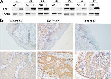
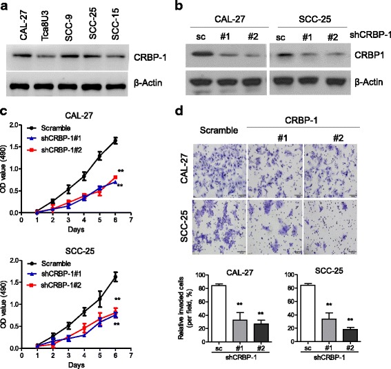
Methods: In this study, an integrated bioinformatics analysis based on the multiple cancer microarray data sets available from Oncomine database was conducted to view the differential expression of CRBP-1 between TSCC and the adjacent non-tumorous tissues. Quantitative real-time polymerase chain reaction (qRT-PCR), western blotting (WB) and immunohistochemical (IHC) assays were performed to investigate CRBP-1 expression in 101 paraffin-embeded TSCC tissues and 48 pairs of freshly frozen tissues. Kaplan-Meier curve and univariate and multivariate Cox-regression analysis were used to estimate the association between CRBP-1 expression and patients' prognosis. Then western blotting, MTT, transwell migration and invasion assays were performed in TSCC cell lines to investigate the effects of CRBP-1 on cellular proliferation and invasion.
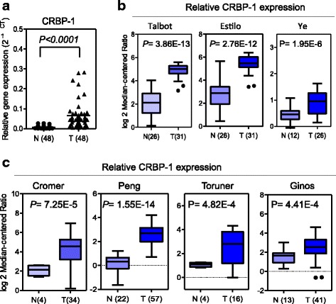
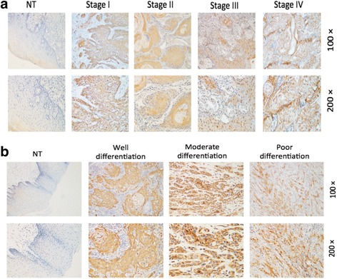
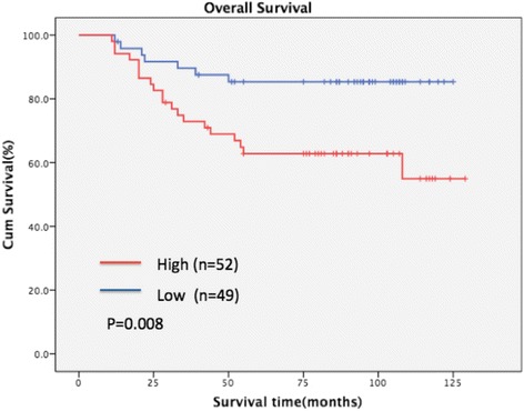
Results: Compared with the matched adjacent non-tumorous tissues, the expression of CRBP-1 was significantly up-regulated in TSCC tissues, which correlated with the differentiation state (P = 0.003), N classification (P = 0.048), the clinical stage (P = 0.048) and death (P = 0.001). The Kaplan-Meier curve showed that TSCC patients with higher CRBP-1 expression levels had lower overall survival rates than those with lower CRBP-1 expression levels. A univariate and multivariate analysis demonstrated that CRBP-1 was an independent prognostic factor (P < 0.05). Furthermore, we knocked down CRBP-1 expression and observed that TSCC cell proliferation and invasion in vitro were significantly blocked, as determined by MTT and transwell assays.
Conclusions: Up-regulated expression of CRBP-1 is associated with poor prognosis in TSCC, so it might potentially serve as an additional prognostic marker, and the inhibition of CRBP-1 might provide new therapeutic approaches for TSCC.
Keywords: CRBP-1; Expression; Invasion; Knockdown; Prognosis; Proliferation; Tongue squamous cell carcinoma.
Conflict of interest statement
Ethics approval and consent to participate
The Ethics Committee of the Sun Yat-sen University Cancer Center approved all the tumor specimens used for this study. All the participating patients in this study have signed the informed consent forms.
Consent for publication
Not applicable.
Competing interests
The authors declare that they have no competing interests.
Publisher’s Note
Springer Nature remains neutral with regard to jurisdictional claims in published maps and institutional affiliations.
Figures
References
-
- Adeel M, Suhail A. Squamous cell carcinoma of oral tongue in young patients – a 10 years tertiary care experience. J Pak Med Assoc. 2016;66:155–158. - PubMed
MeSH terms
Substances
Grants and funding
LinkOut - more resources
Full Text Sources
Other Literature Sources
Research Materials
