. 2018 May 2;20(1):85.
doi: 10.1186/s13075-018-1578-z.

Integrative analysis reveals CD38 as a therapeutic target for plasma cell-rich pre-disease and established rheumatoid arthritis and systemic lupus erythematosus

Affiliations

Integrative analysis reveals CD38 as a therapeutic target for plasma cell-rich pre-disease and established rheumatoid arthritis and systemic lupus erythematosus

Suzanne Cole et al. Arthritis Res Ther. .

Abstract

Background: Plasmablasts and plasma cells play a key role in many autoimmune diseases, such as rheumatoid arthritis (RA) and systemic lupus erythematosus (SLE). This study was undertaken to evaluate the potential of targeting CD38 as a plasma cell/plasmablast depletion mechanism by daratumumab in the treatment of patients with RA and SLE.

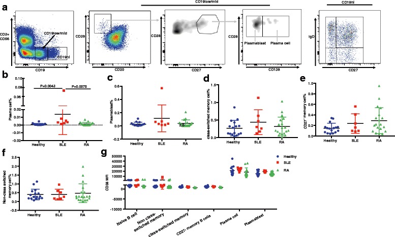
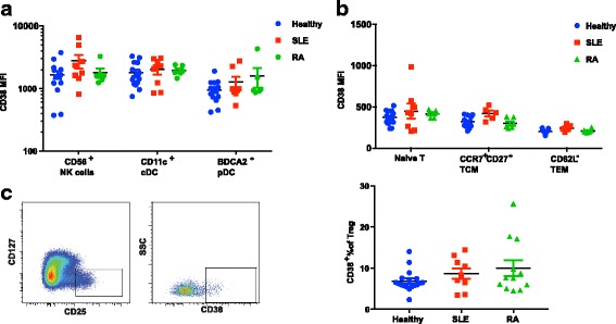
Methods: RNA-sequencing analysis of synovial biopsies from various stages of RA disease progression, flow cytometry analysis of peripheral blood mononuclear cells (PBMC) from patients with RA or SLE and healthy donors, immunohistochemistry assessment (IHC) of synovial biopsies from patients with early RA, and ex vivo immune cell depletion assays using daratumumab (an anti-CD38 monoclonal antibody) were used to assess CD38 as a therapeutic target.

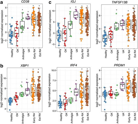
Results: We demonstrated that the plasma cell/plasmablast-related genes CD38, XBP1, IRF4, PRDM1, IGJ and TNFSF13B are significantly up-regulated in synovial biopsies from patients with arthralgia, undifferentiated arthritis (UA), early RA and established RA as compared to healthy controls and control patients with osteoarthritis. In addition, the highest CD38 expression was observed on plasma cells and plasmablasts compared to natural killer (NK) cells, classical dendritic cells (DCs), plasmacytoid DCs (pDCs) and T cells, in blood from healthy controls and patients with SLE and RA. Furthermore, IHC showed CD38 staining in the same region as CD3 and CD138 staining in synovial tissue biopsies from patients with early RA. Most importantly, our data show for the first time that daratumumab effectively depletes plasma cells/plasmablasts in PBMC from patients with SLE and RA in a dose-dependent manner ex vivo.

Conclusion: These results indicate that CD38 may be a potential target for RA disease interception and daratumumab should be evaluated clinically for the treatment of both RA and SLE.

Keywords: CD38; Daratumumab; Plasma cell; Rheumatoid arthritis; Systemic lupus erythematosus.


Conflict of interest statement

Ethics approval and consent to participate

All protocols for collecting synovial biopsies and blood/serum were approved by Institutional Review Board Repatriation General Hospital and Flinders University (Adelaide, Australia), Adelaide Hospital, (Adelaide, Australia), Trinity College Dublin (Dublin, Ireland), St. Vincent’s University Hospital (Dublin, Ireland), and Queen Mary University of London (London, UK). All patients signed the consent form for participating in the study.

Competing interests

SC, AW, XY, JD, MS, CC, HA, AA, IA, LM, SN and YG are current or former employees of Janssen Research & Development, Johnson & Johnson. CP has received research grants and/or honoraria and/or consultation fees from Abbott/AbbVie, Astellas, Astra-Zeneca/MedImmune, BMS, Celgene, Grunenthal, GSK, Janssen/J&J, MSD, Pfizer, Sanofi, Roche/Genentech/Chugai and UCB. MDW, MDS, DJV and UF are recipients of Janssen collaboration research grants. The remaining authors declare that they have no competing interests.

Publisher’s Note

Springer Nature remains neutral with regard to jurisdictional claims in published maps and institutional affiliations.

Figures

Fig. 1
Fig. 1
Plasma cell/plasmablast-related gene expression is increased at different stages of disease progression to rheumatoid arthritis (RA). RNA-sequencing was performed on biopsies from healthy tissue, and tissue from patients with osteoarthritis (OA), arthralgia, undifferentiated arthritis (UA), early RA and established RA (Est RA). a CD38 expression stratified by disease group/stage. b Transcription factors XBP1, IRF4, and PRDM1 mRNA were measured in synovial biopsies from different disease groups. c Plasma cell/plasmablast survival-related genes IGJ and TNFSF13B expression in synovial biopsies from different disease groups. *Statistically significant difference compared to healthy tissue (all graphs) (adjusted P < 0.05)
Fig. 2
Fig. 2
CD38 expression on immune cells in peripheral blood mononuclear cells (PBMC) from healthy donors and patients with systemic lupus erythematosus (SLE) or rheumatoid arthritis (RA). a Gating strategy for plasma cells and plasmablasts in PBMC samples. Plasma cells are gated as live singlet lymphocytes, CD3CD56CD19low/midCD20CD38hiCD27hiCD138+. Plasmablasts are gated as live singlet lymphocytes, CD3CD56CD19low/midCD20CD38hiCD27hiCD138. CD19hi B cells are separated into naïve B cell (IgD+CD27), non-class-switched memory B cell (CD27+IgD+), class-switched memory B cell (CD27+IgD) and CD27 memory B cell (CD27IgD). b Percentage of CD27hiCD38hiCD138+ plasma cells in total lymphocytes from PBMCs from healthy controls and patients with SLE or RA. c Percentage of CD27hiCD38hiCD138 plasmablasts in total lymphocytes from PBMCs from different groups. d-f Percentage of class-switched memory cell (d), CD27 memory B cell (e), and non-class-switched memory B cell (f) in total lymphocytes. g Quantification of CD38 MFI in naïve B cell, non-class switched memory cell, class-switched memory cell, CD27 memory cell, plasma cells and plasmablasts
Fig. 3
Fig. 3
CD38 expression on myeloid, natural killer (NK) and T cells in peripheral blood mononuclear cells (PBMC) from healthy donors and patients with systemic lupus erythematosus (SLE) and rheumatoid arthritis (RA). a Quantification of CD38 MFI on CD56+CD16+ NK cells, CD11c+ classical DC, and BDCA2+ plasmacytoid dendritic cells (DC). b Quantification of CD38 MFI on CD45RA+ naïve T cells, CD62LhiCD27hiCCR7+CD45RA central memory T cells (TCM) and CD62LCD45RA effector memory T cell (TEM). c Representative fluorescence-activated cell sorting (FACS) plot of CD38 expression on CD4+CD127lowCD25hi T regulatory cells (Tregs). d Proportion of the CD38bri subset in peripheral Tregs from healthy controls and donors with SLE or RA
Fig. 4
Fig. 4
Immunohistochemistry analysis (IHC) of CD38 expression in synovial tissue biopsies from patients with early rheumatoid arthritis (RA). a Representative IHC of tissue sections with abundant CD3, CD38 and CD138 staining. b Representative IHC of tissue sections with abundant CD3 but sparse CD38 and CD138 staining. c Representative IHC of tissue sections with rare CD3, CD38 and CD138 staining. All images are shown at × 20 with 100-μM scale bar in each image. Data shown represent synovial biopsies from nine patients with early RA
Fig. 5
Fig. 5
Dose-dependent depletion by daratumumab of plasma cells and plasmablasts from peripheral blood mononuclear cell (PBMC) samples ex vivo. a Representative fluorescence-activated cell sorting (FACS) plot of combined plasma cells/plasmablasts. Shown is pre-gated on live singlet CD3CD56CD19low/midCD20 lymphocytes. From left to right, plot shows the representative plasma cell/plasmablast population at 1 μg/ml isotype control, 0.0003 μg/ml daratumumab, 0.01 μg/ml daratumumab and 1 μg/ml daratumumab (Dara), respectively. Number in the quadrant shows absolute number of CD27hiCD38hi plasma cells-plasmablasts in each condition at 72 h post-culture. b Quantification of plasma cell/plasmablast number at 72 h post-culture with isotype control or daratumumab at indicated concentrations. c Dose-response of plasma cell/plasmablast depletion by daratumumab in all donors with systemic lupus erythematosus (SLE) or rheumatoid arthritis (RA) and healthy donors combined. d Representative FACS plot of CD27IgD memory B cell, CD27+IgD class-switched memory B cell and CD27+IgD+ non-class-switched memory B cell at 72 h post-culture in the presence of daratumumab at each indicated concentration. Number in the quadrant shows absolute number of each memory B cell subset. e-g Slight increase in non-class-switched memory B cell (e), CD27+ class-switched memory B cell (f) and CD27 memory B cell (g) compared to isotype control at each indicated concentration. For each individual donor at each daratumumab concentration, triplicate wells were combined for quantification in a, b and d and then normalized to isotype control in c and e-g Data shown represent four healthy controls, five donors with SLE and four with RA
Fig. 6
Fig. 6
Quantitative PCR analysis shows dose-dependent IGJ down-regulation by daratumumab ex vivo. a Positive correlation between IGJ mRNA and plasma cell count at baseline of isotype control. b Positive correlation between IGJ mRNA and plasma cell count at different daratumumab concentrations ex vivo. c-e IGJ expression at different daratumumab concentration at 72 h post-treatment in healthy controls (c, n = 7) and patients with systemic lupus erythematosus (SLE) (d, n = 4) and rheumatoid arthritis (RA) (e, n = 4). Relative expression of IGJ in the isotype control was normalized to 1 (shown as dashed line). f Positive correlation between normalized IGJ mRNA fold change and plasma cell count fold change compared to isotype control at various daratumumab concentrations. g Correlation between the number of plasma cells at baseline and the maximal IGJ mRNA down-regulation. Data shown in a, b, f and g represent two donors with SLE and sixhealthy control donors

References

    1. Deane KD. Preclinical rheumatoid arthritis (autoantibodies): an updated review. Curr Rheumatol Rep. 2014;16(5):419. doi: 10.1007/s11926-014-0419-6. - DOI - PMC - PubMed
    1. Kurts C, Panzer U, Anders HJ, Rees AJ. The immune system and kidney disease: basic concepts and clinical implications. Nat Rev Immunol. 2013;13(10):738–753. doi: 10.1038/nri3523. - DOI - PubMed
    1. Martin F, Chan AC. B cell immunobiology in disease: evolving concepts from the clinic. Annu Rev Immunol. 2006;24:467–496. doi: 10.1146/annurev.immunol.24.021605.090517. - DOI - PubMed
    1. Jacobi AM, Mei H, Hoyer BF, Mumtaz IM, Thiele K, Radbruch A, Burmester GR, Hiepe F, Dorner T. HLA-DRhigh/CD27high plasmablasts indicate active disease in patients with systemic lupus erythematosus. Ann Rheum Dis. 2010;69(1):305–308. doi: 10.1136/ard.2008.096495. - DOI - PubMed
    1. Nutt SL, Hodgkin PD, Tarlinton DM, Corcoran LM. The generation of antibody-secreting plasma cells. Nat Rev Immunol. 2015;15(3):160–171. doi: 10.1038/nri3795. - DOI - PubMed

MeSH terms